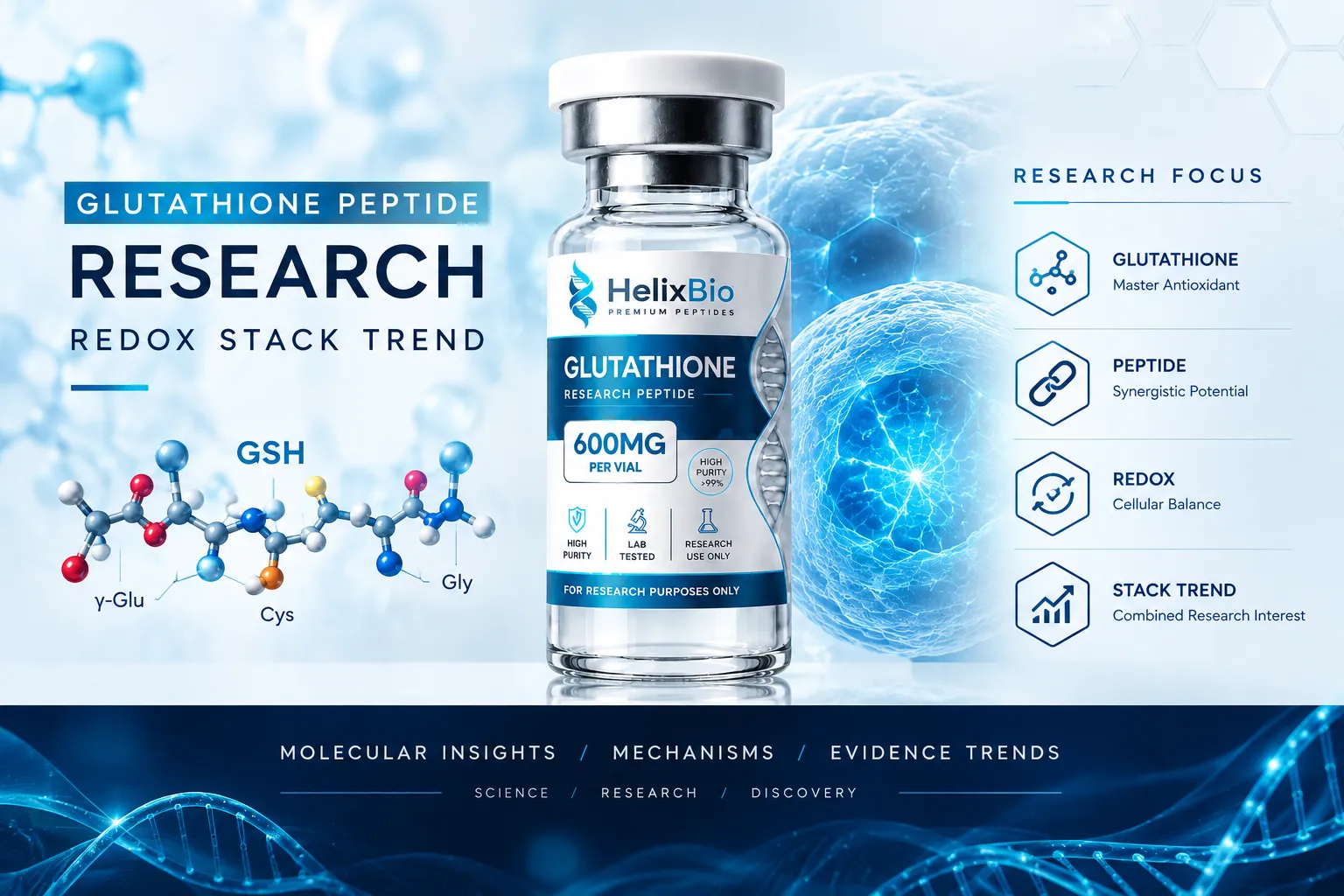
Glutathione is a tripeptide of glutamate, cysteine and glycine joined through an unusual gamma-glutamyl bond that ordinary intracellular peptidases cannot cleave.
Because ribosomes form only alpha peptide bonds, glutathione cannot be made by translation and is instead built in two ATP-dependent enzymatic steps.
GSH is reduced glutathione carrying a free thiol; GSSG is two glutathione molecules joined by a disulfide bond, and the two differ by 305.31 mass units.
Glutathione peroxidase consumes GSH while reducing peroxides, and glutathione reductase regenerates GSH from GSSG using NADPH as the electron source.
NADPH drives glutathione recycling; NAD+ and NADH are a separate redox couple, and nicotinamide nucleotide transhydrogenase is the mitochondrial enzyme that links them.
Glutamate-cysteine ligase is the rate-limiting biosynthetic enzyme and is feedback-inhibited by glutathione, with cysteine as the usual limiting substrate.
Mitochondria hold roughly 10 to 15 percent of cellular glutathione but make none of it; SLC25A39 was identified in 2021 as required for import across the inner membrane.
GSH/GSSG ratios are compartment-, tissue- and method-specific, and artefactual oxidation during sample preparation can inflate measured GSSG substantially.
Glutathione is the most abundant low-molecular-weight thiol in most living cells, and it is genuinely a peptide — three amino acids joined end to end. It is also assembled in a way no ribosome would produce, works through a chemistry that has nothing to do with receptor binding, and is measured by methods that can manufacture the very result they are meant to report. Search interest has recently pulled the term in two directions at once: toward precise biochemistry on one side, and toward combination or "stack" discussions on the other. This article covers the first properly, then uses it to judge the second.
Featured In This Article
Glutathione
RESEARCH PEPTIDE
Highly purified synthetic peptide prepared for rigorous laboratory research.
$72.00
What Is Glutathione, and Why Is It Called a Peptide?
Glutathione is a tripeptide composed of L-glutamate, L-cysteine and glycine, with the molecular formula C10H17N3O6S and a molecular weight of 307.32 g/mol (CAS 70-18-8). Its systematic name, gamma-L-glutamyl-L-cysteinylglycine, records the feature that separates it from an ordinary three-residue peptide.
In a conventional peptide, each bond forms between the alpha-carboxyl group of one residue and the alpha-amino group of the next. In glutathione, the first bond forms instead between the gamma-carboxyl group on the glutamate side chain and the amino group of cysteine. Only the cysteine-glycine bond is a standard alpha peptide bond.
That single structural quirk has two consequences worth stating plainly. The gamma-glutamyl linkage is not recognised by most intracellular peptidases, which is why glutathione survives in the cytosol at millimolar concentrations rather than being degraded within minutes. And because ribosomes can only form alpha peptide bonds, glutathione cannot be made by translation at all. It is built enzymatically, in two ATP-dependent steps, which is why glutathione biosynthesis is a metabolic pathway rather than a gene product.
The thiol is where the chemistry happens
The reactive part of the molecule is the sulfhydryl (-SH) group on the cysteine residue. Glutamate and glycine contribute stability and recognition; cysteine contributes the reactive electron pair. Every redox function attributed to glutathione traces back to that one sulfur atom.
GSH and GSSG: What Actually Differs
GSH is reduced glutathione — the form carrying a free thiol. GSSG is glutathione disulfide, the oxidised form, in which two glutathione molecules are joined through a covalent disulfide bond between their cysteine sulfurs. The abbreviation is worth decoding: in GSH, the G stands for the whole glutathione molecule and SH for the free thiol, so GSSG is literally two G units bridged by S-S.
Property
GSH (reduced)
GSSG (oxidised)
Structure
One tripeptide, free -SH
Two tripeptides joined by -S-S-
Molecular formula
C10H17N3O6S
C20H32N6O12S2
Molecular weight
307.32 g/mol
612.63 g/mol
Redox role
Electron donor; substrate for peroxidases
Electron acceptor; substrate for glutathione reductase
Typical intracellular share
The large majority of the pool
A small minority under normal conditions
Formed by
Biosynthesis; reduction of GSSG
Oxidation of two GSH, enzymatic or spontaneous
The two forms differ by exactly 305.31 mass units, which is not a trivial detail for anyone characterising the material analytically. It means a single mass spectrometry run separates them unambiguously, and it means an oxidised sample is chemically a different substance, not a degraded version of the same one.
How the Glutathione Redox Cycle Works
The common shorthand — glutathione neutralises free radicals — describes one step of a loop and skips the part that makes the system work. The full cycle involves two enzymes and a cofactor.
What glutathione peroxidase does
Glutathione peroxidase (GPx) reduces hydrogen peroxide and organic hydroperoxides to water and the corresponding alcohols, using two molecules of GSH as the electron source and producing one molecule of GSSG. The mammalian GPx1 through GPx4 enzymes are selenoproteins: they carry a selenocysteine residue at the active site, which is why selenium status intersects with glutathione biology at a level most antioxidant writing ignores.
GPx4 deserves separate mention. It is the isoform that reduces phospholipid hydroperoxides within membranes, and its loss triggers ferroptosis, an iron-dependent form of regulated cell death characterised by lipid peroxidation. Because GPx4 requires GSH as its reductant, glutathione depletion and GPx4 inhibition converge on the same cellular outcome. Ferroptosis is one of the reasons glutathione has returned to the centre of current cell-biology research rather than remaining a background housekeeping molecule.
What glutathione reductase does
Glutathione reductase (GR) is the enzyme that closes the loop. It is a flavoenzyme that reduces GSSG back to two molecules of GSH, drawing the electrons from NADPH. Without this step, a cell would consume its glutathione pool during a single sustained oxidative episode. With it, one molecule of glutathione can pass through the cycle repeatedly, and the limiting resource shifts from glutathione itself to the supply of NADPH.
That shift is the single most useful thing to understand about the system. Glutathione status is not only a question of how much glutathione is present. It is also a question of whether the cell can keep regenerating it.
Why NADPH Is Not NAD+
Four cofactors with similar names appear constantly in glutathione discussions, and conflating them produces confident statements that are chemically wrong. They are distinct molecules with distinct roles.
Cofactor
Redox partner
Predominant cellular state
Principal role
NAD+
NADH
Largely oxidised
Accepts electrons in catabolism (glycolysis, TCA cycle, beta-oxidation)
NADH
NAD+
Minority pool
Delivers electrons to the respiratory chain
NADP+
NADPH
Minority pool
Accepts electrons in reductive pathways
NADPH
NADP+
Largely reduced
Supplies reducing power for biosynthesis and antioxidant regeneration
Glutathione reductase uses NADPH. It does not use NADH, and it does not use NAD+. Statements along the lines of "NAD+ recycles glutathione" describe the wrong molecule.
There is, however, a real and specific connection between the two nucleotide pools, and it is more interesting than the loose version. Inside mitochondria, nicotinamide nucleotide transhydrogenase (NNT) sits in the inner membrane and transfers a hydride from NADH to NADP+, using the proton-motive force to drive the reaction forward and maintain a strongly reduced mitochondrial NADPH pool. NNT is therefore a genuine bridge between NADH availability and glutathione recycling — but it is a mitochondrial membrane enzyme dependent on membrane potential, not a general statement about NAD+ supplementation.
A widely used laboratory mouse strain, C57BL/6J, carries a spontaneous inactivating deletion in the Nnt gene. Isolated mitochondria from these animals show a higher GSSG/GSH ratio and impaired peroxide handling compared with Nnt-intact C57BL/6 substrains. A substantial share of the published rodent redox literature was generated in a background missing a major mitochondrial NADPH source, which is worth checking before results are compared across studies.
How Glutathione Is Synthesised and Turned Over
Biosynthesis runs in two ATP-dependent steps in the cytosol.
Glutamate-cysteine ligase (GCL, also called gamma-glutamylcysteine synthetase) joins glutamate and cysteine through the gamma linkage to form gamma-glutamylcysteine. This is the rate-limiting step.
Glutathione synthetase adds glycine to form glutathione.
Two control features matter for interpreting research. GCL is feedback-inhibited by GSH itself, so the pathway self-limits as the pool fills, which is why simply supplying more precursor does not scale glutathione indefinitely. And cysteine is normally the limiting substrate, because it is the least abundant of the three amino acids and is consumed by competing pathways. That is the entire mechanistic basis for interest in cysteine-delivery strategies, and it is the correct place to introduce NAC rather than treating NAC as a form of glutathione.
Turnover runs through the gamma-glutamyl cycle. Because intracellular peptidases cannot cleave the gamma bond, degradation requires gamma-glutamyl transferase (GGT), an enzyme on the outer surface of the plasma membrane. Glutathione must therefore be exported before it can be broken down, and its constituent amino acids are recovered and re-imported. Extracellular glutathione is largely a substrate for this cycle rather than a molecule that enters cells intact.
Mitochondrial Glutathione: A Pool With No Factory
Mitochondria generate a large share of cellular reactive oxygen species and hold roughly 10 to 15 percent of total cellular glutathione. They also contain none of the biosynthetic machinery. Both GCL and glutathione synthetase are cytosolic, so every molecule of mitochondrial glutathione has to cross the inner mitochondrial membrane — and glutathione is anionic at physiological pH, so it does not diffuse across.
How it got there was an open question for decades. In 2021, Wang and colleagues identified SLC25A39, a mitochondrial carrier of previously unknown function, as required for glutathione import into the matrix. Loss of SLC25A39 reduced mitochondrial glutathione without changing whole-cell glutathione levels, and cells lacking both SLC25A39 and its paralogue SLC25A40 showed defects in iron-sulfur cluster proteins. Subsequent work has described how the transporter is regulated, including its degradation by the matrix protease AFG3L2 and its stabilisation under low matrix glutathione.
Two things follow. First, whole-cell glutathione measurements can be uninformative about the compartment where oxidative chemistry is most concentrated, since the two pools are regulated separately. Second, compartmentalisation is now a tractable experimental variable rather than an acknowledged limitation, which is the main reason mitochondrial redox has become one of the more active areas of current glutathione research.
What the GSH/GSSG Ratio Can and Cannot Tell You
The GSH/GSSG ratio is frequently presented as a universal readout of oxidative status, as though a single number described a whole organism. It does not.
The ratio is compartment-specific: cytosolic, mitochondrial, nuclear and extracellular glutathione exist at different concentrations and different redox states within the same cell at the same moment. It is tissue-specific. It is also, importantly, method-specific. The half-cell reduction potential of the GSSG/2GSH couple depends on the square of the GSH concentration, not on the ratio alone, so two samples with identical ratios at different absolute concentrations are not in equivalent redox states.
Then there is the measurement problem. GSH autoxidises to GSSG readily once a sample is removed from its biological context, and because GSSG is normally present at a very low concentration, even small artefactual oxidation inflates it disproportionately. Reported micromolar GSSG concentrations in whole blood have been attributed to sample preparation rather than to biology. Reliable protocols block the thiol at the moment of collection, typically with N-ethylmaleimide, which both prevents autoxidation and inhibits glutathione reductase.
When comparing GSH/GSSG figures between published studies, check whether thiol-blocking was performed at collection and which derivatising agent was used. A ratio obtained without in-situ derivatisation is not directly comparable with one obtained with it.
How Glutathione Is Characterised in the Laboratory
Analytical questions about glutathione as a research material are narrower than the biology, and they follow directly from the thiol.
Identity is established by mass, most commonly by LC-MS or LC-MS/MS. The 305.31 unit gap between GSH and GSSG makes oxidation state visible in the same run.
Purity is typically reported by HPLC area percent. Detection near 210 to 215 nm is required, because glutathione contains no tryptophan or tyrosine and therefore has no useful absorbance at 280 nm.
Oxidation state is the specific issue this molecule raises. An area-percent purity figure is only meaningful if the chromatographic method actually resolves GSSG from GSH. If it does not, oxidised material can be counted inside the main peak.
Sample handling governs whether any of the above stays true. Thiols oxidise on exposure to air, elevated pH and trace metal ions.
A certificate of analysis documents what was measured on one batch, on one date, by one method. It can establish identity, purity by a stated method, and — where an appropriate assay was run — content. It does not establish biological activity, efficacy, or safety in any organism, and it says nothing about a different lot. Those are separate categories of evidence and should not be substituted for one another.
What "Glutathione Stack" Means, and Where the Evidence Sits
The phrase glutathione stack is community terminology, not a scientific category. It describes combining glutathione with other compounds selected because they appear in the same metabolic diagrams. The term has appeared more frequently in search data recently, though the underlying query volumes are small and the growth figures come from low or unstated baselines, so the signal is better read as emerging vocabulary than as established demand.
The useful question is not which combinations are popular but which have direct evidence, which have a mechanistic rationale only, and which have neither. Those are different claims.
Combination
Mechanistic rationale
Direct combination evidence
Interpretation
Glutathione + NAC
Strong: NAC supplies cysteine, the rate-limiting substrate for GCL
Limited; most data concern NAC alone, not the pair
Precursor and product, not equivalents
Glycine + NAC (GlyNAC)
Strong: supplies both the limiting substrate and the second amino acid
One small randomised trial in older adults
Best-characterised combination, but small
Glutathione + NAD+
Indirect: NADPH drives GSSG reduction; NNT links NADH to NADPH in mitochondria
None identified for co-administration
Distinct redox couples; do not describe as interchangeable
Glutathione + vitamin C
Plausible: ascorbate and glutathione interact in coupled redox chemistry
Glutathione and NAC are not the same claim
N-acetylcysteine is an acetylated form of cysteine. It is not glutathione, does not contain glutamate or glycine, and has no gamma-glutamyl bond. Its relationship to glutathione is upstream: it supplies the substrate that limits the first synthetic step. Evidence generated with NAC therefore supports statements about cysteine availability and about NAC, and transferring it to glutathione as a molecule is an unjustified step. The reverse transfer is equally unjustified.
GlyNAC — glycine plus NAC — is the most rigorously studied of the combinations discussed under the stack heading. A randomised, placebo-controlled trial in older adults reported improvements in glutathione concentrations, oxidative stress markers, mitochondrial fuel oxidation and several functional measures over 16 weeks. The finding is real and the design was controlled, but the trial randomised twelve participants per arm. That is a pilot-scale result, and it is routinely cited as though it were a definitive one.
Glutathione and NAD+ occupy different parts of the same map
Interest in pairing glutathione with NAD+ precursors comes from the observation that both appear in mitochondrial energy diagrams. The accurate mechanistic statement is narrower: glutathione recycling depends on NADPH; NADPH and NAD+ are different molecules; and NNT provides a membrane-potential-dependent link between the mitochondrial NADH and NADPH pools. No published study establishing a co-administration effect was identified. The two systems are related, not redundant. Helix Bio's separate overview of NAD+ mechanism and the clinical trial record on NAD+ precursors covers that side in detail, and the general question of how combinations are assessed is treated in the mechanism-based peptide stacking guide.
What the Evidence Establishes and What Remains Open
Established at the level of biochemistry and cell biology: glutathione's structure and gamma linkage, the GSH/GSSG couple, the catalytic roles of glutathione peroxidase and reductase, NADPH dependence of recycling, the two-step biosynthetic pathway with GCL as rate-limiting, GGT-dependent turnover, and SLC25A39-dependent mitochondrial import. These are not contested.
Well supported but context-dependent: the association between altered glutathione status and a long list of disease states. Association is the accurate word. Depleted glutathione is a consequence of oxidative processes as often as a cause of them, and most human data are observational.
Open or thin: whether raising glutathione by any route produces a defined biological outcome; how much of an administered dose survives GGT-mediated extracellular processing; whether specific combinations outperform their components; and how well any measured pool represents the compartments doing the chemistry. Mechanistic importance is not the same as demonstrated efficacy, and a molecule can be indispensable to cellular function without being a useful intervention.
Materials described on this page are supplied for laboratory research only. Nothing here is a protocol, a dosing recommendation, or a statement about human use.
Got Questions?
Frequently Asked Questions
Glutathione is a tripeptide, meaning a peptide of three amino acid residues: L-glutamate, L-cysteine and glycine. The term glutathione peptide is used in research catalogues to distinguish the intact tripeptide from its individual amino acid components and from cysteine-donor compounds such as N-acetylcysteine.
The bond between glutamate and cysteine forms at the gamma-carboxyl group of the glutamate side chain rather than at its alpha-carboxyl group. Ribosomes can only form alpha peptide bonds, so glutathione is assembled enzymatically instead of being translated, and most intracellular peptidases cannot cleave the gamma linkage.
GSH is reduced glutathione, carrying a free sulfhydryl group on its cysteine residue. GSSG is glutathione disulfide, in which two glutathione molecules are covalently joined through a disulfide bond between those sulfur atoms. GSH is the electron donor; GSSG is the oxidised product formed when it donates.
Glutathione peroxidase reduces hydrogen peroxide and organic hydroperoxides, oxidising two molecules of GSH to one molecule of GSSG in the process. The mammalian GPx1 to GPx4 enzymes are selenoproteins carrying a selenocysteine residue at the active site, which makes selenium status relevant to glutathione-dependent peroxide handling.
Glutathione reductase is a flavoenzyme that reduces GSSG back to two molecules of GSH, drawing electrons from NADPH. This recycling step is what allows a fixed glutathione pool to handle repeated oxidative challenges, and it makes NADPH supply a limiting factor in sustained glutathione function.
NADPH is the reducing cofactor used by glutathione reductase to regenerate GSH from GSSG. Glutathione recycling therefore depends directly on NADPH availability. NADPH is distinct from NADH, and NADP+ is distinct from NAD+, despite the similarity of the abbreviations.
No. Glutathione reductase uses NADPH, not NAD+ or NADH. A genuine link exists inside mitochondria, where nicotinamide nucleotide transhydrogenase transfers a hydride from NADH to NADP+ using the proton-motive force, but that is a membrane-potential-dependent enzyme rather than a general statement about the NAD+ pool.
Glutamate-cysteine ligase joins glutamate and cysteine through the gamma linkage to form gamma-glutamylcysteine, then glutathione synthetase adds glycine. Both steps consume ATP and occur in the cytosol. The first step is rate-limiting and is feedback-inhibited by glutathione itself.
No. N-acetylcysteine is an acetylated form of the single amino acid cysteine. It contains no glutamate, no glycine and no gamma-glutamyl bond. Its relationship to glutathione is as an upstream substrate supply, so research findings for one should not be transferred to the other.
It gives a relative measure of oxidation within one sampled compartment, analysed by one method. Interpretation depends on tissue, subcellular compartment, absolute concentrations and assay technique, so ratios are not comparable across studies unless the sampling and derivatisation procedures match.
Mitochondria generate a large share of cellular reactive oxygen species and hold roughly 10 to 15 percent of cellular glutathione, but contain none of the biosynthetic enzymes. Glutathione must be imported across the inner membrane, and the two pools are regulated separately, so whole-cell measurements may not reflect mitochondrial status.
A certificate documents identity, purity by a stated method, and where measured, content, for one specific batch on one date. For a thiol compound the analytical method must resolve GSSG from GSH for the purity figure to be meaningful. A certificate does not establish biological activity, efficacy or safety.