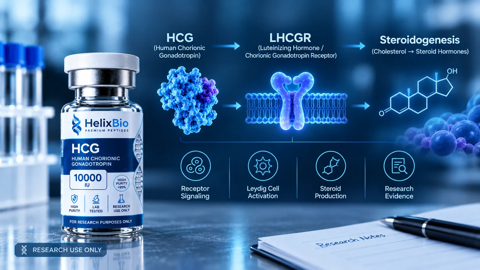
HCG is a heterodimeric glycoprotein hormone whose alpha subunit is shared with LH, FSH and TSH and whose beta subunit confers receptor specificity.
HCG binds LHCGR, a Gs-coupled G protein-coupled receptor expressed on Leydig cells and on ovarian theca, granulosa and luteal cells.
Receptor binding, receptor activation, intracellular transduction and steroidogenic output are four separately measured claims, not one.
Single-molecule work published in 2026 resolved two distinct hCG binding mechanisms within the LHCGR extracellular domain, with binding lifetimes of 18.4 and 0.67 seconds.
PKA signalling induces StAR, which transports cholesterol into the mitochondrion and constitutes the rate-limiting step in steroid synthesis.
In COS-7 cells expressing human LHCGR, cAMP half-maximal effective concentrations were about 107 pM for hCG and about 530 pM for LH, a roughly five-fold difference.
LH reaches its maximal effect faster than hCG and, in human granulosa cells, preferentially engages ERK1/2 and AKT rather than the steroidogenic arm.
In mouse Leydig cells the two hormones differed in early signalling but produced comparable CREB phosphorylation, Stard1 expression and testosterone output.
The widely repeated claim that hCG is ten times more potent than LH with equal testosterone output combines two studies in different species and cell types.
Clinical evidence for approved chorionic gonadotropin products is specific to those products and does not extend to research-grade material.
Human chorionic gonadotropin acts on the same receptor as luteinizing hormone. That single fact generates more interpretive error in commercial writing about the hormone than anything else in its biology: a shared receptor gets read as shared behaviour, and shared behaviour gets read as transferable evidence. The published record disagrees, and it disagrees with numbers attached. What follows traces the chain from hCG binding the LH/CG receptor (LHCGR) through Gs, cAMP and PKA to the steroidogenic enzymes, then sets out where hCG and LH measurably separate and what that separation means for reading a study.
Featured In This Article
HCG
RESEARCH PEPTIDE
Highly purified synthetic peptide prepared for rigorous laboratory research.
$72.00
The short version
hCG is a heterodimeric glycoprotein hormone. Its alpha subunit is shared with LH, FSH and TSH; its beta subunit carries a C-terminal extension that no other member of the family has. It binds LHCGR, a Gs-coupled G protein-coupled receptor expressed on Leydig cells in the testis and on theca, granulosa and luteal cells in the ovary. Receptor occupancy raises intracellular cAMP, activates protein kinase A, and drives a transcriptional programme whose best-characterised outputs are StAR and the cytochrome P450 steroidogenic enzymes.
Every arrow in that paragraph is a separate experimental claim with its own evidence. The structural and analytical layer — subunit lengths, glycosylated mass, glycoform heterogeneity, why gonadotropin potency is expressed in International Units rather than milligrams — is not repeated here. This page is about the signalling chain and the evidence attached to it.
Binding, activation and downstream effect are three claims, not one
Vendor descriptions routinely compress the whole cascade into a single sentence: hCG binds LHCGR and raises testosterone. That sentence skips four independently measured steps, each established by a different method and each with its own failure modes.
Stage
Molecular event
What a positive result establishes
Typical evidence
Binding
hCG occupies the LHCGR extracellular domain
Ligand and receptor interact
Radioligand binding, force spectroscopy, structural work
Activation
Receptor conformation changes and couples Gs
The interaction is productive, not merely adhesive
cAMP accumulation, BRET and FRET conformational sensors
StAR and P450 enzymes are induced, steroid appears
The cascade reaches a measurable biological endpoint
Gene expression, steroid quantification
Binding itself is not even one event. Single-molecule force spectroscopy published in 2026 resolved two distinct binding mechanisms within the LHCGR extracellular domain — one associated with the leucine-rich repeats, one with the hinge loop — separated by more than an order of magnitude in binding lifetime, 18.4 seconds against 0.67 seconds. The same dual mode appeared on full-length receptor in living cells. A single dissociation constant quoted for hCG at LHCGR describes an average over at least two physically different interactions.
From receptor to cAMP
LHCGR belongs to the glycoprotein hormone receptor family and carries an unusually large N-terminal extracellular domain: eleven leucine-rich repeats that do the hormone binding, a structurally complex hinge region, and a conventional seven-transmembrane bundle. Hormone binding at the ectodomain has to be converted into a conformational change at the membrane, which is why the hinge region attracts as much attention as the binding site itself.
Once Gs is engaged, the route is the textbook one. The stimulatory G protein activates adenylyl cyclase, intracellular cAMP rises, cAMP-dependent protein kinase A is released from its regulatory subunits, and PKA phosphorylates cytoplasmic targets and the transcription factor CREB. The pathway is not unique to gonadotropins — the GHRH receptor runs the same Gs-cAMP route in the pituitary, which is set out in the GHRH analogues versus GHRPs comparison — and that generic quality is worth holding onto. What makes an LHCGR experiment specific is the receptor, the cell type and the endpoint, not the second messenger.
Steroidogenesis: what PKA switches on
The rate-limiting step in steroid synthesis is not an enzyme. It is the delivery of cholesterol from the outer to the inner mitochondrial membrane, and that transfer depends on steroidogenic acute regulatory protein (StAR), a short-lived protein whose expression responds rapidly to cAMP-PKA signalling. Once cholesterol arrives, CYP11A1 cleaves its side chain to give pregnenolone, and the pathway then branches by cell type.
In Leydig cells, pregnenolone passes through CYP17A1 and the hydroxysteroid dehydrogenases to testosterone. In the ovary, theca cells generate androgen precursors that granulosa cells aromatise to estradiol through CYP19A1 under FSH control, and luteal cells convert pregnenolone to progesterone through 3-beta-HSD. The FDA-approved labelling for chorionic gonadotropin injection describes exactly this division: stimulation of the interstitial cells of the testis to produce androgens, and of the corpus luteum to produce progesterone.
The branch point matters for experimental design. The same ligand at the same receptor produces a different steroid depending on which enzymes the cell expresses. A result obtained in a Leydig line is a statement about testosterone biosynthesis in that line, not about ovarian steroidogenesis, and the reverse holds too. The granulosa case carries an extra complication: aromatisation depends on CYP19A1 expression that is itself driven by FSH, so an estradiol endpoint in that compartment reports on a two-hormone system rather than on LHCGR activation alone.
Attribute findings to the ligand actually used. A large share of the literature on LHCGR signalling was generated with LH, and a large share of the writing about hCG cites it. The two are different molecules with different receptor residence times, and the evidence below shows they do not produce identical outputs even in the same cell.
hCG versus LH: the measured differences
The comparison has been done directly. Using recombinant hormones in COS-7 cells stably expressing human LHCGR, hCG was roughly five-fold more potent than LH for cAMP production: half-maximal effect at 107.1 plus or minus 14.3 pM for hCG against 530.0 plus or minus 51.2 pM for LH. Potency was not the only difference. LH reached its maximal effect faster, within about ten minutes against roughly an hour for hCG, and in primary human granulosa-lutein cells held at equipotent doses for up to 36 hours, cAMP production oscillated and ran significantly higher with hCG.
Those two observations together create a design problem that is easy to miss. If potency and kinetics both differ, then a comparison at matched molar concentration and a comparison at matched timepoint will each mis-rank the hormones, in opposite directions. The studies that found qualitative differences got there by dosing to equal effect rather than equal concentration, and by sampling across a time course. A paper that does neither is not evidence that the two hormones behave the same.
The divergence is qualitative as well as quantitative. In those human granulosa cells, hCG drove the cAMP-PKA-steroidogenic arm while LH preferentially engaged ERK1/2 and AKT — a pattern later characterised with BRET and FRET sensors as biased agonism at LHCGR. Work published in 2025 proposes receptor trafficking as the mechanism: differential handling of the occupied receptor after internalisation directs which signal predominates.
Property
hCG
LH
Why it matters when reading a study
cAMP potency at human LHCGR
ED50 about 107 pM
ED50 about 530 pM
Equipotent dosing, not equal dosing, is required for a fair comparison
Kinetics of maximal response
Slower, about one hour
Faster, about ten minutes
Single-timepoint experiments can invert the apparent ranking
Preferred downstream arm in human granulosa cells
cAMP, PKA, steroidogenesis
ERK1/2 and AKT
The hormones support different cellular programmes, not different amounts of one
Receptor residence
Longer, attributed to the beta-subunit C-terminal extension
Shorter
Where the "ten times more potent" claim comes from
A figure circulating across vendor pages states that hCG is about ten times more potent than LH while producing equivalent testosterone output. That sentence welds two separate results from two different experimental systems. The potency figure traces to transfected COS-7 cells and is five-fold, not ten. The equal-testosterone finding comes from a different study in mouse Leydig cells, where the same group reported that the rodent receptor mediates a quantitatively but not qualitatively different response. Species, cell type, receptor construct, endpoint and assay all differ between the two. Neither reports a ten-fold cAMP ratio, and the combined claim is not a property of the hormone.
What human evidence exists, and what it is evidence about
Approved prescription products containing chorionic gonadotropin exist in the United States, with labelled indications covering prepubertal cryptorchidism not due to anatomical obstruction, hypogonadotropic hypogonadism in males, and induction of ovulation in selected anovulatory infertility. The approved labelling also carries a statement, required since 1975, that chorionic gonadotropin has no known effect on fat mobilisation, appetite or body fat distribution, and has not been demonstrated to be effective adjunctive therapy in the treatment of obesity.
That body of evidence is real and it is narrow. Drug approval attaches to a named product from a named manufacturer at a defined strength, administered by a defined route, for a defined indication, using material characterised to pharmacopoeial standards and assayed for potency in International Units. None of that transfers to a research material. Published clinical work on pharmaceutical hCG establishes nothing about the identity, potency, purity or suitability of any laboratory preparation, including Helix Bio's HCG research material, and no responsible page should imply otherwise.
Because hCG is a glycosylated heterodimer rather than a single-sequence peptide, a chromatographic area-percent purity figure does not carry the meaning it carries on a synthetic peptide certificate. Check which method produced the number and whether assayed potency and related-gonadotropin limits are reported at all. The [certificate of analysis guide](/how-to-read-peptide-certificate-of-analysis) covers the general reading; the glycoprotein-specific caveats sit with the product documentation.
What recent work has added
Three strands from 2025 and 2026 genuinely move the picture rather than restate it. The force-spectroscopy result described above splits hCG binding into two mechanisms with very different lifetimes. The trafficking work offers a mechanistic account of why two agonists at one receptor produce different signals, moving the explanation past a simple affinity argument. And a 2026 review of LHCGR pharmacodynamics during follicular maturation consolidates the evolutionary, genetic and physiological case that LH and hCG occupy separate roles rather than substituting for one another.
A fourth strand is structural and unresolved. Every human glycoprotein hormone receptor structure determined by cryo-electron microscopy so far has been monomeric, while functional evidence has long pointed to these receptors operating as dimers; a 2025 structure of an invertebrate leucine-rich-repeat receptor, an evolutionary predecessor of the family, was reported as a dimer. Whether the signalling unit at LHCGR is one receptor or two is therefore not settled, and that is a live question rather than a detail.
None of this is a change in clinical practice, and none of it is a finding about any commercially supplied research material. It is a sharpening of the mechanistic picture, which is the level at which the hormone is actually used as a reagent.
What the evidence does not establish
Receptor-level potency does not predict biological output. The clearest demonstration is the rodent Leydig result, where measurably different early signalling converged on the same testosterone.
In vitro work in transfected cells establishes what a receptor construct does in that system. It does not establish tissue behaviour, and receptor density in a transfected line is not physiological.
Findings generated with LH are not findings about hCG, and findings in mouse cells are not findings in human cells. The same laboratory reached different conclusions about qualitative divergence depending on which it used.
Approved-product evidence is evidence about that product. It does not extend to research-grade material of different source, preparation and formulation.
Signalling bias has been characterised in a small number of cell systems. Whether the same bias operates across every LHCGR-expressing tissue has not been established.
The oligomeric state of the signalling receptor is not resolved. Structural and functional lines of evidence currently disagree, so any model drawn as a single receptor plus a single G protein is a simplification.
Nothing in the receptor literature speaks to safety, effectiveness or suitability for any use in humans, and nothing here should be read as supporting any such use.
Research-use context
hCG is useful in the laboratory for a specific reason: it is a well-characterised ligand at a receptor that also has a second natural agonist, which makes it a good system for asking how ligand properties shape a signalling outcome when the receptor is held constant. That question sits within a wider reproductive axis — kisspeptin and GnRH upstream of the gonadotropins, covered in the kisspeptin and KISS1R research guide — and the axis framing is often more informative than treating any one hormone in isolation.
Material supplied for research is labelled for research use only and is not intended for human or veterinary administration by any route. The general position is set out in the guide to research-use-only compliance in the United States.
Got Questions?
Frequently Asked Questions
HCG (human chorionic gonadotropin) is a heterodimeric glycoprotein hormone produced by the placental syncytiotrophoblast. It consists of an alpha subunit shared with LH, FSH and TSH and a beta subunit unique to hCG, associated non-covalently, with a substantial fraction of the molecule's mass contributed by attached carbohydrate. In laboratory research it is used principally as a ligand at the LH/CG receptor.
hCG binds LHCGR, the luteinizing hormone/choriogonadotropin receptor, a Gs-coupled G protein-coupled receptor. LHCGR is expressed on Leydig cells in the testis and on theca, granulosa and luteal cells in the ovary. It is the same receptor that luteinizing hormone activates, which is why the two hormones are frequently and incorrectly treated as interchangeable.
LHCGR is a glycoprotein hormone receptor with a large N-terminal extracellular domain containing eleven leucine-rich repeats, a structurally complex hinge region, and a seven-transmembrane bundle. Hormone binding occurs at the extracellular domain and must be transmitted through the hinge to the transmembrane region to produce G protein coupling.
Receptor activation engages Gs, which stimulates adenylyl cyclase and raises intracellular cAMP, activating protein kinase A and leading to CREB phosphorylation. The key downstream target is StAR, which transports cholesterol into the mitochondrion — the rate-limiting step. CYP11A1 then converts cholesterol to pregnenolone, after which cell-type-specific enzymes determine whether the product is testosterone, androgen precursors, estradiol or progesterone.
Binding means the hormone occupies the receptor; activation means the occupied receptor changes conformation and couples to a G protein. They are measured differently and can be dissociated experimentally. Single-molecule work published in 2026 found two distinct binding mechanisms within the LHCGR extracellular domain with binding lifetimes of 18.4 and 0.67 seconds, so even binding is not a single uniform event.
They share an identical alpha subunit and act at the same receptor, but the hCG beta subunit carries a C-terminal extension that LH's does not, which is the accepted explanation for its longer persistence. Functionally, hCG is more potent for cAMP at human LHCGR while LH acts faster and, in human granulosa cells, preferentially engages ERK1/2 and AKT rather than the steroidogenic arm.
No published measurement identified supports that ratio. In COS-7 cells expressing human LHCGR, the cAMP half-maximal effective concentrations were approximately 107 pM for hCG and 530 pM for LH, a roughly five-fold difference. The widely circulated ten-fold figure, usually paired with a claim of equivalent testosterone output, combines results from two different studies in two different species and cell types.
The beta subunit confers receptor specificity. All four human glycoprotein hormones share the same alpha subunit, so what distinguishes hCG from LH, FSH and TSH is beta. In hCG the beta subunit additionally carries a carboxy-terminal peptide extension bearing O-linked oligosaccharides, a feature absent from LH beta and associated with hCG's longer circulating survival.
This is the phenomenon described as biased agonism, characterised for hCG and LH at LHCGR using BRET and FRET approaches. Work published in 2025 proposes receptor trafficking as a mechanistic explanation: differential handling of the occupied receptor after internalisation influences which downstream arm predominates. The account is mechanistic and remains an active research question rather than a settled one.
Approved prescription products containing chorionic gonadotropin carry labelled indications in prepubertal cryptorchidism not due to anatomical obstruction, hypogonadotropic hypogonadism in males, and induction of ovulation in selected anovulatory infertility. That evidence was generated with characterised pharmaceutical preparations at defined strengths by defined routes and is specific to those products.
The status depends on the product, not the molecule. FDA-approved chorionic gonadotropin drug products exist for the indications above, and their labelling has been required since 1975 to state that the hormone has no known effect on fat mobilisation, appetite or body fat distribution and has not been demonstrated effective in treating obesity. Research-grade material is not an approved drug, has not been evaluated for safety or effectiveness, and is supplied for laboratory use only.
No. Approval and clinical evidence attach to a specific product from a specific manufacturer at a specific strength, characterised to pharmacopoeial standards and assayed for potency in International Units. A research preparation of different source, formulation and documentation is a different material, and the published clinical literature establishes nothing about its identity, potency or suitability.