L-Carnitine Research: The Carnitine Shuttle and the Evidence
Metabolic researchSeptember 13, 202612 min read
L-Carnitine moves long-chain fatty acids into mitochondria through a four-step shuttle. The mechanism explained, and what human evidence does and does not show.
L-Carnitine is a quaternary ammonium betaine (C7H15NO3, 161.20 g/mol) and contains no peptide bond, despite being biosynthesised from lysine and methionine.
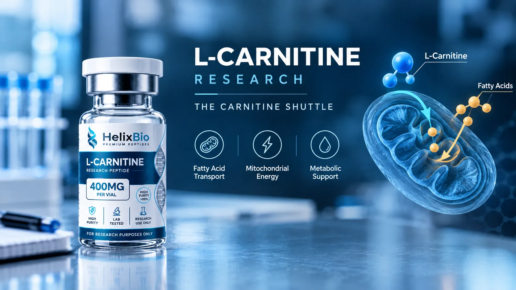
The carnitine shuttle is a four-step cycle: acyl-CoA synthetase activates the fatty acid, CPT1 transfers the acyl group to carnitine, CACT translocates the acylcarnitine, and CPT2 regenerates acyl-CoA inside the matrix.
CPT1 sits in the outer mitochondrial membrane, exists as three tissue isoforms, and is inhibited by malonyl-CoA, making it the regulated control point of long-chain fatty acid entry.
CACT (SLC25A20) is the only component that physically crosses the inner mitochondrial membrane, operating as an electroneutral one-to-one acylcarnitine/free-carnitine antiport.
CPT2 is a single ubiquitously expressed enzyme on the matrix face of the inner membrane and is not regulated by malonyl-CoA, which makes it biologically distinct from CPT1.
Carnitine is regenerated rather than consumed by the shuttle, and it does not itself carry out beta-oxidation; transport capacity and oxidation rate are separate variables.
In a 24-week randomised study in 14 healthy men, oral L-carnitine with carbohydrate raised muscle total carnitine by about 21 percent and altered fuel selection during exercise, but later work found no change in adaptations to high-intensity interval training.
A meta-analysis of 37 randomised controlled trials found a modest pooled body weight reduction of roughly 1.2 kg, with only the body weight effect persisting when analysis was restricted to high-quality trials.
Acylcarnitine profiles by tandem mass spectrometry report chain-length-specific species and are an established readout of fatty acid oxidation in research and newborn screening.
Conventional HPLC purity does not distinguish L-carnitine from the racemate, so enantiomeric purity requires a chiral method or specific optical rotation.
L-Carnitine earns its place in metabolic research for one structural reason: long-chain fatty acids cannot cross the inner mitochondrial membrane on their own. A dedicated transport system moves them, and L-Carnitine is the carrier that system runs on. This article works through that system enzyme by enzyme, then separates what the biochemistry firmly establishes from what human trials have and have not shown. The mechanism is settled science. The clinical picture is not, and the gap between the two is where most published descriptions of this compound go wrong.
Featured In This Article
L-Carnitine
RESEARCH PEPTIDE
Highly purified synthetic peptide prepared for rigorous laboratory research.
$25.00
L-Carnitine Spray
RESEARCH PEPTIDE
Highly purified synthetic peptide prepared for rigorous laboratory research.
$89.99
L-Carnitine Is a Small Molecule, Not a Peptide
L-Carnitine is a quaternary ammonium betaine with the molecular formula C7H15NO3 and a molecular weight of 161.20 g/mol. It contains no peptide bond. The molecule is biosynthesised from lysine and methionine, which is the likely origin of the recurring error that files it alongside peptides, but a biosynthetic precursor is not a structural classification. L-Carnitine belongs to the same practical category as other small molecules that turn up in peptide catalogues, such as 5-Amino-1MQ, which is also not a peptide.
At physiological pH the molecule is a zwitterion. The quaternary nitrogen carries a permanent positive charge and the carboxylate a negative one. That permanent charge is the reason carnitine cannot diffuse freely across a lipid bilayer and depends on the OCTN2 transporter to enter cells — a constraint that shapes every experiment involving carnitine availability.
Only the L-enantiomer, pharmacopoeially named levocarnitine, is biologically active. The D-form is not. Full identity data, including CAS number, InChIKey and monograph status, sits on the L-Carnitine Spray product listing.
The Carnitine Shuttle in Four Steps
The carnitine shuttle is often described in one sentence, which hides the fact that it is a cycle with four distinct chemical events across two membranes. Stated precisely:
Two features of this cycle drive most of its research use. First, carnitine is regenerated rather than consumed — it is a shuttle, not a substrate. Second, each transfer produces an acylcarnitine whose chain length reflects the fatty acid involved, and those species are stable enough to measure directly.
CPT1 Is the Regulated Step
CPT1 sits in the outer mitochondrial membrane with its catalytic domain facing the cytosol. It catalyses the transfer of an acyl group from coenzyme A to the hydroxyl of carnitine, producing an acylcarnitine and releasing free CoA. This is conventionally described as the rate-limiting step of long-chain fatty acid oxidation, and the description is defensible: everything downstream depends on flux through it.
Three isoforms, three tissue contexts
CPT1 exists as three isoforms. CPT1A is the dominant liver form, CPT1B the muscle and heart form, and CPT1C is expressed in brain. The isoforms differ in kinetics and in sensitivity to inhibition, which is why a result obtained in hepatocytes does not transfer cleanly to a muscle preparation. Experimental designs that manipulate CPT1 need to name the isoform.
Malonyl-CoA is the control signal
CPT1 is inhibited by malonyl-CoA, the first committed intermediate of fatty acid synthesis. The logic is a reciprocal switch: when a cell is building fatty acids, malonyl-CoA rises and entry of fatty acids into the mitochondrion for degradation is suppressed. Malonyl-CoA concentration is in turn set by acetyl-CoA carboxylase, so the shuttle is coupled to the cell's wider anabolic state rather than simply to substrate availability. CPT1B is markedly more sensitive to malonyl-CoA inhibition than CPT1A, another reason the isoform distinction is not cosmetic.
CACT Is the Step Most Explanations Skip
Carnitine-acylcarnitine translocase, encoded by SLC25A20, occupies the inner mitochondrial membrane and is routinely omitted from summaries that name only CPT1 and CPT2. Omitting it makes the shuttle incoherent, because neither CPT enzyme moves anything across the inner membrane.
CACT operates as an antiporter. It imports one acylcarnitine into the matrix while exporting one free carnitine outward, in an electroneutral one-to-one exchange that requires no additional energy input. That exchange stoichiometry is what makes the cycle self-sustaining: the free carnitine released by CPT2 inside the matrix is the counter-substrate that drives the next import. A recent review of carnitine metabolism in kidney disease states this exchange explicitly, alongside the observation that CACT dysfunction produces acylcarnitine accumulation and impaired beta-oxidation.
CPT2 Reverses the Reaction Inside the Matrix
CPT2 is bound to the matrix face of the inner mitochondrial membrane and catalyses the reverse of the CPT1 reaction: it transfers the acyl group from carnitine back onto matrix coenzyme A. The products are an acyl-CoA molecule positioned where beta-oxidation enzymes can act on it, and a free carnitine that becomes CACT's export substrate.
Unlike CPT1, CPT2 is a single ubiquitously expressed enzyme with no tissue-specific isoforms, and it is not regulated by malonyl-CoA. The two enzymes catalyse chemically related reactions in opposite directions, under different constraints, on opposite sides of a membrane.
Component
Membrane and orientation
Function
Key distinction
CPT1
Outer membrane, catalytic site facing cytosol
Acyl-CoA to acylcarnitine
Three tissue isoforms; inhibited by malonyl-CoA; regulatory control point
CACT (SLC25A20)
Inner membrane
Acylcarnitine in, free carnitine out
The only step that physically crosses the inner membrane; electroneutral 1:1 antiport
CPT2
Inner membrane, matrix face
Acylcarnitine back to acyl-CoA
Single ubiquitous form; no malonyl-CoA regulation; supplies beta-oxidation directly
A useful test of any carnitine shuttle description: if it names an enzyme that moves fatty acids across the inner mitochondrial membrane, it is wrong. CPT1 and CPT2 are acyltransferases. Only CACT translocates.
Transport Is Not Oxidation
The shuttle delivers substrate. It does not oxidise anything. Once acyl-CoA is regenerated in the matrix, beta-oxidation proceeds through its own repeating four-reaction cycle — dehydrogenation, hydration, a second dehydrogenation, and thiolytic cleavage — shortening the chain by two carbons per pass and releasing acetyl-CoA, which enters the TCA cycle.
This distinction matters because it bounds what carnitine availability can do. Increasing carnitine raises the capacity of a transport step. Whether that translates into greater fatty acid oxidation depends on whether transport was limiting in the first place, and on the capacity of the beta-oxidation and TCA machinery downstream. In most healthy tissue under most conditions, it is not obvious that transport is the binding constraint. L-Carnitine does not perform beta-oxidation, and a statement that it "burns fat" collapses a transport function into a metabolic outcome that has to be demonstrated separately.
Free Carnitine, Acylcarnitines and the Carnitine Pool
The total carnitine in a tissue or a sample is distributed between unesterified free carnitine and a spectrum of acylcarnitines. Together these constitute the carnitine pool, and its composition is informative in a way the total is not.
Species
Notation
What it is
Research relevance
Free carnitine
C0
Unesterified carnitine available for CPT1
Denominator of the acyl-to-free ratio; falls when acyl groups accumulate
Short-chain acylcarnitines
C2, C3, C4, C5
Acetyl- and other short acyl groups
Reflects matrix acetyl-CoA load and branched-chain amino acid flux
Medium-chain acylcarnitines
C6 to C12
Medium-chain acyl groups
Rises in specific oxidation defects
Long-chain acylcarnitines
C14 to C18
The species the shuttle transports
Accumulates when CPT2 or CACT function is impaired
Because each species is chain-length specific and stable, tandem mass spectrometry of acylcarnitine profiles is an established readout of fatty acid oxidation — it underpins newborn screening for oxidation disorders and is a common endpoint in metabolic research. The pattern locates the lesion: long-chain species accumulating upstream of a blocked reconversion step look different from a general carnitine shortage in which free carnitine itself is low.
What Human Studies Actually Show
The evidence base for L-Carnitine is layered, and the layers are frequently merged in secondary sources. Kept apart, it looks like this.
Evidence type
What it addresses
Strength
Principal limitation
Biochemical and structural
Shuttle mechanism, enzyme function, transport stoichiometry
Established
Says nothing about whether altering carnitine changes an outcome
Genetic deficiency states
CPT1, CPT2, CACT and OCTN2 defects
Well characterised
Describes pathology of absence, not effects of addition in intact systems
Animal and cell models
Mechanistic manipulation of shuttle components
Informative
Species and model differences; not human outcomes
Human controlled trials
Muscle metabolism, body composition, metabolic markers
Mixed and modest
Muscle metabolism and exercise
The controlling problem in this literature is that raising muscle carnitine content is difficult. In a 24-week randomised, double-blind study in 14 healthy men, pairing oral L-carnitine with carbohydrate raised muscle total carnitine by roughly 21 percent, while a carbohydrate-only control showed no change. At low exercise intensity the carnitine group used substantially less muscle glycogen, consistent with greater lipid use; at high intensity the metabolic picture shifted differently, with higher pyruvate dehydrogenase activation and lower muscle lactate. Work output in the performance trial rose about 11 percent from baseline.
That study is frequently cited as settling the question. It should not be. It was small, single-sex, and required 24 weeks and an insulin stimulus to move muscle carnitine at all. Work from the same research group later found that increasing skeletal muscle carnitine availability did not alter the adaptations produced by high-intensity interval training. The honest summary is that carnitine loading can measurably shift fuel selection in human muscle under specific conditions, and that this has not reliably converted into training or performance benefit.
Body weight and composition
A meta-analysis of 37 randomised controlled trials reported a statistically significant but small reduction in body weight, on the order of about 1.2 kg, with reductions in BMI and fat mass and no significant effect on waist circumference or body fat percentage. When the analysis was restricted to high-quality trials, only the body weight effect held. The effect was most apparent in adults with overweight or obesity.
A significant pooled effect of roughly a kilogram, attenuating when trial quality is filtered, is not the result the mechanism is usually invoked to support. It is a real finding and a modest one.
What Researchers Are Studying Now
Current activity has moved away from performance questions and toward the shuttle's role in metabolic disease, where the interesting signal is dysfunction rather than supplementation.
A 2026 review in the Journal of Translational Medicine maps carnitine dysregulation in diabetic kidney disease as a connected sequence: impaired OCTN2-mediated carnitine reabsorption in renal tubular epithelial cells under high-glucose conditions, suppression of CPT1 and CPT2 activity by insulin resistance and reactive oxygen species, and consequent intracellular accumulation of long-chain acylcarnitines, which the authors link to tubular injury pathways. The same review describes a stage-dependent pattern in the carnitine pool, with reduced free carnitine and elevated specific acylcarnitine species in blood. Separately, work published in JCI Insight examined impaired carnitine-induced fatty acid oxidation in both experimental and human diabetic kidney disease, reporting downregulation of CPT1A and CPT2 in kidneys from patients with diabetes.
Two cautions are worth attaching to this line of work. It is largely mechanistic, observational and model-based rather than interventional, so it identifies the shuttle as a candidate target rather than establishing that modifying it changes outcomes. And a cluster of recent publications on a topic is a research direction, not a consensus.
What This Evidence Does Not Establish
Specific to L-Carnitine, the following do not follow from anything above:
That increased transport capacity produces increased fat oxidation in a given tissue. Transport is limiting only where it is limiting.
That a demonstrated shift in fuel selection during exercise produces a training, performance or body-composition outcome. The one human study that raised muscle carnitine and altered fuel use is not matched by consistent downstream benefit.
That findings for acetyl-L-carnitine or propionyl-L-carnitine apply to free L-Carnitine. These are distinct esters with separate literatures.
That deficiency-state clinical data transfer to non-deficient systems. Evidence supporting levocarnitine in defined carnitine-deficiency indications describes a different question entirely.
That CPT1 and CPT2 changes observed in diseased kidney tissue predict the effect of carnitine administration in that tissue.
That a Certificate of Analysis speaks to efficacy. Analytical documentation characterises a batch of material and nothing more.
Laboratory Characterisation and Its Limits
Two analytical questions dominate carnitine work, and neither is answered by a single number.
Identity is established by mass spectrometry against the known molecular formula, supported by chromatographic retention against a reference standard. Carnitine has both USP and European Pharmacopoeia monographs, so a result can be checked against a public standard rather than a supplier claim.
Purity by conventional HPLC reports chemical purity — it does not distinguish L-carnitine from the racemate, because enantiomers are chemically identical to a non-chiral method and to a mass spectrometer. Enantiomeric purity requires a chiral method or specific optical rotation. A second recurring problem is mass basis: because free-base carnitine is markedly hygroscopic, material is often supplied as tartrate, fumarate or hydrochloride, and a stated milligram figure may describe the salt rather than the carnitine within it.
An analytical certificate documents properties of one batch on one date by one set of methods. It establishes nothing about biological activity, human safety, therapeutic suitability or clinical effect. Interpreting one is a separate skill, covered in the [guide to reading a Certificate of Analysis](/how-to-read-peptide-certificate-of-analysis).
Acetyl-L-carnitine and propionyl-L-carnitine are esters of carnitine carrying an acetyl or propionyl group respectively, with different molecular formulas and different physical properties. Both occur endogenously as short-chain acylcarnitines produced by normal shuttle activity, so their presence in a sample is expected. What does not follow is the transfer of findings between them. Each has developed its own experimental literature, and a study reporting an effect of one ester provides no evidence about free L-Carnitine unless the study measured it.
Got Questions?
Frequently Asked Questions
L-Carnitine is a small quaternary ammonium betaine with the molecular formula C7H15NO3 and a molecular weight of 161.20 g/mol. Its established biological function is transport: it carries long-chain fatty acids across the inner mitochondrial membrane so they can undergo beta-oxidation. It is biosynthesised from lysine and methionine and also obtained from the diet.
No. Peptides are chains of amino acids joined by peptide bonds, and L-Carnitine contains no peptide bond. It is a single small molecule classed as a betaine. The confusion usually arises because lysine and methionine are its biosynthetic precursors, but a precursor relationship is not a structural classification.
The carnitine shuttle is the cycle that moves long-chain fatty acids into the mitochondrial matrix. CPT1 transfers an acyl group from coenzyme A onto carnitine, CACT exchanges the resulting acylcarnitine across the inner membrane for free carnitine, and CPT2 regenerates acyl-CoA inside the matrix. Carnitine is recycled rather than consumed.
CPT1 sits in the outer mitochondrial membrane and catalyses the transfer of an acyl group from acyl-CoA to carnitine, producing an acylcarnitine. It is treated as rate-limiting because all downstream long-chain fatty acid oxidation depends on flux through it, and because it is the step subject to regulation by malonyl-CoA.
Malonyl-CoA, the first committed intermediate of fatty acid synthesis, inhibits CPT1. This creates a reciprocal switch: when a cell is synthesising fatty acids, entry of fatty acids into the mitochondrion for degradation is suppressed. The muscle isoform CPT1B is more sensitive to this inhibition than the liver isoform CPT1A.
They catalyse chemically related reactions in opposite directions on opposite sides of a membrane. CPT1 is in the outer membrane facing the cytosol, exists as three tissue isoforms, and is malonyl-CoA regulated. CPT2 is on the matrix face of the inner membrane, is a single ubiquitously expressed enzyme, is not malonyl-CoA regulated, and reconverts acylcarnitine back to acyl-CoA.
Carnitine-acylcarnitine translocase, encoded by SLC25A20, is the inner mitochondrial membrane transporter that moves acylcarnitine into the matrix while exporting free carnitine outward in an electroneutral one-to-one exchange. It is the only component of the shuttle that physically translocates anything across the inner membrane, which is why descriptions naming only CPT1 and CPT2 are incomplete.
The shuttle delivers substrate to beta-oxidation but is not part of it. Beta-oxidation is a separate repeating cycle of reactions in the matrix that shortens the acyl chain by two carbons per pass and releases acetyl-CoA. L-Carnitine does not perform beta-oxidation; it makes the substrate available to the enzymes that do.
Free carnitine, denoted C0, is unesterified and available as a CPT1 substrate. Acylcarnitines are carnitine esterified to acyl groups of varying chain length, produced during transport. Together they form the carnitine pool, and the distribution between them is often more informative in research than the total figure.
Findings are mixed. One 24-week randomised study in 14 healthy men raised muscle total carnitine by about 21 percent when L-carnitine was paired with carbohydrate, and measurably shifted fuel selection during exercise. Subsequent work from the same group found that increasing muscle carnitine availability did not alter adaptations to high-intensity interval training.
That phrasing conflates a transport role with a metabolic outcome. Mechanistically L-Carnitine enables long-chain fatty acid entry into mitochondria, but whether raising carnitine increases oxidation depends on whether transport was limiting. A meta-analysis of 37 randomised trials reported a modest pooled body weight reduction of roughly 1.2 kg, with only that effect persisting among high-quality trials.
It can document identity, chemical purity, appearance and related properties for one batch on one date using stated methods. It cannot establish biological activity, human safety or therapeutic value. Two carnitine-specific limits also apply: conventional HPLC purity does not separate L-carnitine from the racemate, and a stated milligram figure may refer to a salt form rather than carnitine content.