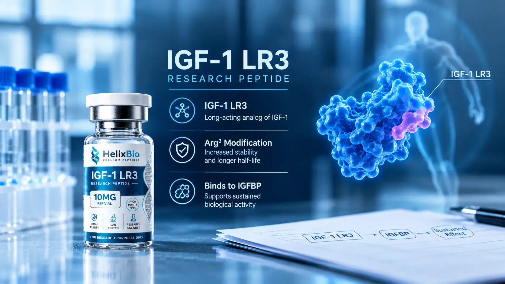
IGF-1 LR3 is an 83-residue recombinant analogue of human IGF-1, differing from the native 70-residue hormone by an Arg3 substitution and a 13-residue N-terminal extension.
The Arg3 substitution and the N-terminal extension were characterised separately in 1992, so each modification's contribution is traceable to primary sources.
No primary source establishing the widely quoted 100-fold or 1,000-fold IGFBP affinity reduction as a general property of the molecule could be located.
Recombinant porcine IGFBP-3 suppresses IGF-I-stimulated differentiation of L6 myogenic cells but does not suppress Long-R3-IGF-I-stimulated differentiation.
Receptor affinity has been shown not to predict proliferative effect across IGF-I variants, so binding, activation and functional response are separate claims.
Long [R3] IGF-I reduced average daily gain and feed intake in pigs, reversing the direction of effect reported in rat models.
The commonly cited 20 to 30 hour half-life traces to a mouse atherosclerosis study that delivered the analogue by osmotic minipump and reports no pharmacokinetic measurements.
No clinical trial of IGF-1 LR3 was identified in registry searches conducted in September 2026; every trial located under IGF-1 terms used native recombinant human IGF-1 or mecasermin.
Long R3 IGF-1, written IGF-1 LR3 across most research catalogues, is one of the better-characterised IGF-1 analogues in the laboratory literature and one of the worst-described in the commercial literature. The molecule itself is not in dispute. What is in dispute is nearly every number attached to it. Three figures appear on almost every page selling this compound: that it binds IGF binding proteins 100 to 1,000 times more weakly than native IGF-1, that it is two to three times more potent, and that it has a half-life of 20 to 30 hours. This article traces each of those to its source, and separates what has been measured from what has been inferred. What follows is the evidence layer rather than the molecular specification.
Featured In This Article
IGF-1 LR3
RESEARCH PEPTIDE
Highly purified synthetic peptide prepared for rigorous laboratory research.
$88.00
The two modifications, and which one does the work
Native IGF-1 is a 70-residue single-chain polypeptide of roughly 7,649 daltons, folded by three intramolecular disulfide bonds. IGF-1 LR3 changes it twice. An arginine replaces the glutamate at position 3. A thirteen-residue extension is added to the N-terminus. The result is 83 residues and approximately 9,111 daltons.
A reasonable question follows: which change is responsible for the reduced binding-protein affinity?
The Adelaide group that produced these analogues asked exactly that, and the answer sits in the title of the paper. Francis and colleagues published a panel of recombinant fusion protein analogues in 1992 under the heading "Novel recombinant fusion protein analogues of insulin-like growth factor (IGF)-I indicate the relative importance of IGF-binding protein and receptor binding for enhanced biological potency." A companion paper by King and colleagues in the same volume characterised the Arg3 substitution on its own, in material carrying no extension.
That is a factorial design. The substitution and the extension were separated and compared rather than tested as a single package. Researchers writing about what Arg3 contributes have a primary source available, and it is almost never cited.
What that literature supports is that both changes reduce binding-protein affinity, and that the combination reduces it further than either alone. What it does not support, and what is asserted constantly, is a tidy structural account of charge reversal breaking specific salt bridges inside an IGFBP binding cleft. Glutamate is acidic and arginine is basic, so the substitution does reverse charge at that position. But a plausible interpretation of a measured affinity change is not a demonstrated structural mechanism, and the distinction matters when the interpretation is repeated as though a structure had been solved.
What reduced IGFBP binding does and does not mean
The functional claim is easier to support than the numerical one.
There is a clean cell-model demonstration. Xi and colleagues, working with L6 myogenic cells, showed that recombinant porcine IGFBP-3 suppresses IGF-I-stimulated differentiation but does not suppress Long-R3-IGF-I-stimulated differentiation. That is the property shown functionally, in a named cell line, against a named binding protein.
The fold-change figures are a different matter. Claims of 100-fold and 1,000-fold reductions appear across vendor pages and aggregator entries with no attached measurement. No primary source establishing either figure as a general property of the molecule could be located. There is also a structural reason to distrust any single number: there are six IGF binding proteins, affinity is measured by different methods under different conditions, and a value obtained for IGFBP-3 in one assay is not a value for IGFBP-1 in another.
The accurate formulation is narrower and more useful. Long R3 IGF-1 binds IGF binding proteins considerably more weakly than native IGF-1 does, and in specific systems a specific binding protein fails to inhibit it. That describes measured interactions, not an abolition of binding, and the phrase "IGFBP-independent" overstates it.
Reduced affinity is not independence. IGF-1 LR3 binds IGF binding proteins; it binds them less well than native IGF-1 does. Research framing should reflect that difference, because a fold-change figure quoted for one binding protein in one assay does not describe the whole IGFBP family.
Binding, activation and response are three different claims
Commercial content routinely collapses these. The molecule binds IGF1R, therefore it activates IGF1R, therefore it produces a response, therefore the response scales with affinity. Each step is a separate empirical question.
The type 1 IGF receptor is a receptor tyrosine kinase. Ligand engagement drives receptor autophosphorylation, IRS-1 recruitment, and signalling through the PI3K/AKT and MAPK/ERK cascades. That receptor biology is well established, and studies using IGF-1 LR3 as a ligand describe what the receptor does when engaged by it.
The inference from affinity to outcome is where the trouble lies, and there is direct evidence against it within the IGF-I literature. Jansson and colleagues measured binding kinetics of IGF-I fusion proteins to IGFBP-1 and to a soluble IGF type I receptor by biosensor analysis, then measured the growth-promoting effect of the same proteins on SaOS-2 cells. The affinities did not track the proliferative effect. The fusion with the lowest receptor affinity matched native IGF-I for proliferative potential, while a variant with twice that affinity produced roughly 70 per cent of its activity. The authors concluded that determinants other than receptor affinity regulate IGF-I proliferative action.
That study used Z-domain fusions rather than the LR3 analogue, so it is not an LR3 result. It is a caution at the level of the ligand class, and it is sufficient to make "retains most of its receptor affinity, therefore comparable activity" an unsafe sentence to write.
What has been measured, and in what model
Level
Representative source
What it establishes
Boundary
Biochemical
Francis 1992; King 1992
Both modifications reduce IGFBP affinity; relative contributions compared
Binding measurements only
Receptor kinetics
Jansson 1997
Receptor affinity does not predict proliferative effect
Z-domain fusions, not LR3
Cell culture
Xi 2004
IGFBP-3 fails to suppress LR3-stimulated differentiation
One cell line, one binding protein
Animal, rat
Tomas 1992
Analogue outperformed native IGF-I in a catabolic rat model
Source of the "3x potency" figure
The effect reverses between species
The most consistent omission across pages selling this compound is that Long R3 IGF-1 is not uniformly anabolic across animals.
Dunaiski and colleagues infused Long [R3] IGF-I into finisher pigs at 180 micrograms per kilogram per day for four days. Average daily gain fell. Feed intake fell. Plasma IGFBP-3, IGF-I and insulin concentrations all fell. The paper's own framing is explicit: analogues that bind poorly to IGF binding proteins stimulate growth in the rat and, in contrast, inhibit growth in the pig.
Conlon and colleagues found something adjacent in guinea pigs. A seven-day infusion raised the fractional weight of adrenals, gut, kidneys and spleen, but body weight gain, feed intake, feed conversion efficiency and carcass composition were not significantly affected. Plasma IGF-I and IGF-II both fell, apparently through a reduction in circulating binding proteins.
Two independent groups, two species, and in neither case the straightforward growth effect the rat model produced. A molecule engineered to escape binding-protein control also perturbs the binding-protein system it escapes, and what that produces downstream depends on the animal. Any statement about what IGF-1 LR3 does needs a species attached to it.
Where the circulating numbers came from
The three times more potent figure
This traces to Tomas and colleagues, 1992, in Biochemical Journal, under the title "Insulin-like growth factor-I (IGF-I) and especially IGF-I variants are anabolic in dexamethasone-treated rats."
The comparison is real and the paper is sound. But it is a rat study, in a glucocorticoid-induced catabolic state, measuring nitrogen balance and body-weight endpoints. "Approximately three times more potent than IGF-1" is a result obtained in that model. It is not a molecular potency constant, it is not a human figure, and read alongside the pig data it is not a species-general one either.
The 20 to 30 hour half-life figure
This one does not survive inspection.
The claim is widely sourced to von der Thusen and colleagues, 2011, in American Journal of Pathology. That paper is titled "IGF-1 has plaque-stabilizing effects in atherosclerosis by altering vascular smooth muscle cell phenotype." It studies atherosclerotic plaque phenotype in apolipoprotein E knockout mice. Long R3 IGF-1 was delivered by subcutaneous osmotic minipump, and the reported measurements are plaque morphometry, vascular smooth muscle cell content, intraplaque haemorrhage rate, and plasma IGF-1 and IGFBP-3 by ELISA.
It is not a pharmacokinetic study. It reports no elimination half-life. A continuous osmotic minipump is, if anything, the delivery method chosen when steady exposure cannot be assumed from a molecule's own persistence.
There is a second problem. The tertiary source most responsible for circulating this figure states 20 to 30 hours in its text while listing 56 to 72 hours in its own summary box, citing different sources for each. Two incompatible half-life figures, on one page, for one molecule, and neither is a human measurement.
The defensible position is that no human pharmacokinetic data for IGF-1 LR3 were identified, and that any half-life quoted for it is preclinical or inferred. The separate and frequently confused distinction between a molecule's half-life and a material's shelf-life is covered in peptide half-life vs shelf-life.
Two versions of the extension sequence
This matters for analytical verification. Two versions of the thirteen-residue N-terminal extension circulate. One carries a serine-serine pair, the other a leucine-leucine pair. The same widely-read reference page prints one form in its prose and the other in its full sequence listing.
The serine-serine form is the one that appears in published full-sequence listings and in reference-supplier documentation. A researcher checking a supplied sequence against a secondary source can land on the wrong string depending on which paragraph is read.
Human evidence
No clinical trial of IGF-1 LR3 was identified in registry searches conducted for this article in September 2026.
That statement needs its boundaries stated alongside it. Searches of ClinicalTrials.gov under IGF-1 terms return a substantial body of trials, and every one located concerns native recombinant human IGF-1 or mecasermin: rhIGF-1 in primary IGF-1 deficiency, an rhIGF-1/rhIGFBP-3 complex in retinopathy of prematurity, rhIGF-1 in cystic fibrosis, rhIGF-1 in PAPP-A2 deficiency, and rhGH/rhIGF-1 combination therapy. None used the LR3 analogue. This is a negative finding from the registries searched on a stated date, not proof that no such study exists anywhere.
The mecasermin point deserves emphasis, because it is the most consequential confusion in this space. A recombinant human IGF-1 product is approved in the United States. Its active ingredient is native 70-residue IGF-1, sequence-identical to the endogenous hormone. It is a different molecule from IGF-1 LR3 by thirteen residues and one substitution, and its approval, labelling, clinical evidence and safety database do not transfer.
What this does not establish
The "three times more potent" figure is a dexamethasone-treated rat result, not a molecular potency constant and not a human figure.
No human pharmacokinetic data were identified, so no half-life figure in circulation is a human measurement.
Reduced IGFBP affinity and documented non-inhibition by one binding protein in specific assays is not IGFBP independence.
Binding, receptor activation and functional response are separate measurements, and affinity has been shown not to predict proliferative effect across IGF-I variants.
Two non-rodent species showed no overall growth effect or a reversed one, so no general growth claim is supported.
Evidence for native IGF-1 or mecasermin does not transfer to this analogue.
A certificate of analysis documents attributes of a specific lot. For a recombinant protein folded by three disulfide bonds, an incorrectly paired isomer contains the same atoms in the same number as a correct one, so its mass is identical and it may co-elute chromatographically. Chromatographic purity and mass confirmation therefore cannot report on folding. The measurement that does is a functional potency assay with a stated ED50. See [how to read a peptide certificate of analysis](/how-to-read-peptide-certificate-of-analysis).
Research-use and regulatory position
IGF-1 LR3 is not an FDA-approved drug and is not approved for any indication. The approved recombinant human IGF-1 product is a different molecule, as above.
Under the World Anti-Doping Code, IGF-1 and its analogues fall within Section S2, prohibited at all times, in and out of competition. The United States Anti-Doping Agency states that all forms of exogenous IGF-1 are prohibited at all times, and that any substance containing any form of exogenous IGF-1 should be treated as prohibited. IGF-1 LR3 is an IGF-1 analogue and falls within that class.
It is also worth placing the compound correctly among the growth-axis research peptides it is often shelved beside. IGF-1 and its analogues are neither GHRH analogues nor growth hormone secretagogues, as set out in GHRH analogues vs GHRPs. IGF-1 LR3 acts at the IGF-1 receptor directly and does not stimulate growth hormone release.
Regulatory status should not be inferred from commercial availability, from laboratory use, or from the status of related molecules in the IGF family. The general position on research-use-only classification in the United States is covered in are research peptides legal in the USA.
Helix Bio supplies IGF-1 LR3 as a research material for qualified laboratory work. It is not intended for human or veterinary use, self-administration, or administration by any route.
Got Questions?
Frequently Asked Questions
IGF-1 LR3 is an 83-residue recombinant analogue of human insulin-like growth factor 1. It differs from the native 70-residue hormone by two changes: arginine replaces glutamate at position 3, and a thirteen-residue extension is added to the N-terminus. Together these substantially reduce its affinity for IGF binding proteins.
Long Arginine 3. The arginine refers to the substitution at position 3, and the long refers to the N-terminal extension. The two halves of the name describe the two modifications, which is why material carrying only one of them is a different compound.
Both do, and the combination reduces affinity further than either alone. Analogues separating the two were published in 1992, so this is answerable from primary sources rather than inference. The frequently repeated charge-reversal explanation is a plausible interpretation of the affinity data, not a demonstrated structural mechanism.
No primary source establishing either figure as a general property could be located. There are six IGF binding proteins, and affinity varies by protein, method and condition, so a single universal multiplier is unlikely to be meaningful. What is well supported is considerably weaker binding overall, plus a functional demonstration that IGFBP-3 fails to suppress Long-R3-IGF-I-stimulated differentiation in L6 myogenic cells.
No, and the mix-up is consequential in research models. R3 IGF-I carries only the position-3 substitution and is 70 residues. IGF-1 LR3 carries that substitution plus the thirteen-residue extension, making it 83 residues. They differ by roughly 1,460 daltons, so a single mass-spectrometry run separates them.
They modify the native molecule in opposite directions. DES(1-3)IGF-1 removes the first three N-terminal residues, leaving 67. IGF-1 LR3 adds thirteen and substitutes one, giving 83. Both reduce binding-protein affinity, which is why they are often grouped, but findings on one do not transfer to the other.
No human pharmacokinetic data were identified. The commonly quoted 20 to 30 hour figure is usually sourced to a 2011 mouse atherosclerosis study that delivered the analogue by osmotic minipump and reports no pharmacokinetic measurements at all. A second figure of 56 to 72 hours circulates from the same tertiary source, and neither is a human measurement.
The approximately threefold figure comes from a 1992 study in dexamethasone-treated rats. It is a result in one species in one induced catabolic state, not a molecular potency constant. Read against pig data showing reduced growth, it is not species-general either.
It acts as a ligand at the type 1 IGF receptor, a receptor tyrosine kinase signalling through IRS-1 to the PI3K/AKT and MAPK/ERK cascades. That pathway biology is a property of the receptor. Studies using this analogue describe what the receptor does when engaged, which is not the same as establishing an outcome in an organism.
No clinical trial of IGF-1 LR3 was identified in registry searches conducted in September 2026. Every trial located under IGF-1 search terms used native recombinant human IGF-1 or mecasermin. This is a negative finding from the sources searched on a stated date rather than proof that no such study exists.
No. A recombinant human IGF-1 product is approved in the United States, but its active ingredient is native 70-residue IGF-1, a different molecule. That approval, its labelling and its clinical evidence do not extend to this analogue.
Because it is a recombinant protein folded by three intramolecular disulfide bonds, and an incorrectly paired isomer contains the same atoms in the same number as a correct one. Its mass is identical, so mass spectrometry cannot separate them, and it may co-elute chromatographically. The measurement that reports on folding is a functional potency assay with a stated ED50.