KPV is the C-terminal tripeptide of alpha-MSH. What its sequence and mass actually are, which mechanisms are demonstrated, and where the evidence stops.
KPV is a tripeptide with the sequence lysine-proline-valine, formula C16H30N4O4, molecular weight 342.44 g/mol, CAS 67727-97-3 and PubChem CID 125672.
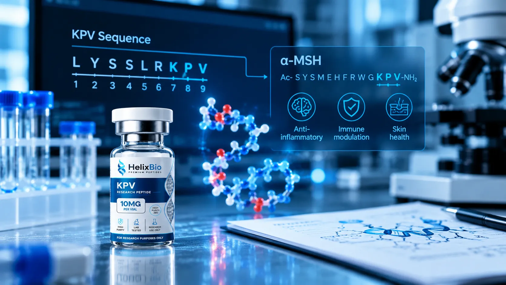
KPV corresponds to residues 11-13 of alpha-MSH, the C-terminal end of Ac-Ser-Tyr-Ser-Met-Glu-His-Phe-Arg-Trp-Gly-Lys-Pro-Val-NH2.
Inside alpha-MSH the terminal valine is amidated, while isolated KPV is supplied as the free acid, so the two forms differ by approximately one dalton.
KPV does not contain the His-Phe-Arg-Trp core that melanocortin receptor binding depends on, and it reduced mortality in MC1R-deficient mice with DSS colitis.
Dalmasso and colleagues showed in Gastroenterology in 2008 that KPV enters cells through the di- and tripeptide transporter PepT1, encoded by SLC15A1, with nanomolar concentrations inhibiting NF-kB and MAP kinase activation.
The proposed importin-alpha3 mechanism rests on a single in vitro dot blot experiment and is described in conditional terms by its own author.
KPV contains no aromatic residues, so absorbance at 280 nm cannot measure its concentration and HPLC purity work must run near 214 nm.
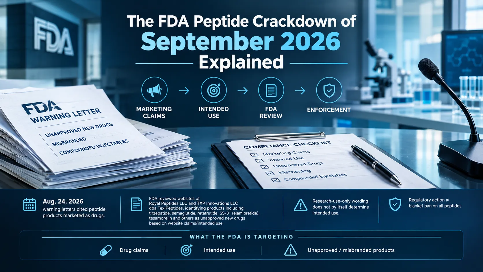
FDA reviewers identified no human safety studies for KPV in the July 2026 PCAC briefing documents; the committee voted 8-6 to recommend inclusion anyway, a non-binding vote that creates no human evidence.
Ask a researcher what KPV is and most will answer correctly in six words: the C-terminal tripeptide of alpha-MSH. That answer is right, and it is also where nearly every published description stops. What sits underneath it is more interesting — what the molecule actually is on a certificate of analysis, which parts of its mechanism have been demonstrated and which are still proposed, and exactly where the evidence stops. KPV is among the smallest compounds in the research peptide catalogue, and that size makes almost every question about it unusually concrete. A three-residue molecule has nowhere to hide.
Featured In This Article
KPV
RESEARCH PEPTIDE
Highly purified synthetic peptide prepared for rigorous laboratory research.
$77.00
What KPV Is
KPV is a tripeptide with the sequence lysine-proline-valine. In single-letter amino acid code that is K-P-V, which is where the name comes from; in three-letter code, Lys-Pro-Val. Its molecular formula is C16H30N4O4, its molecular weight is 342.44 g/mol, and it is catalogued under CAS 67727-97-3 and PubChem CID 125672.
Three residues is very small for a bioactive compound. For scale, BPC-157 has fifteen and thymosin beta-4 has forty-three. KPV weighs roughly a quarter of what BPC-157 does and about a fourteenth of what full-length thymosin beta-4 does. That has consequences the literature keeps running into: KPV is short enough to be a substrate for peptide transporters rather than a ligand for a receptor, short enough to be degraded quickly by peptidases, and short enough that the usual analytical shortcuts for peptides do not work on it.
The Sequence and What "Alpha-MSH(11-13)" Really Means
Alpha-melanocyte-stimulating hormone is a tridecapeptide — thirteen residues — cleaved from the precursor protein proopiomelanocortin. Its full sequence is Ac-Ser-Tyr-Ser-Met-Glu-His-Phe-Arg-Trp-Gly-Lys-Pro-Val-NH2. Number those residues one through thirteen and the last three are lysine, proline, valine. That is why KPV is written in the literature as alpha-MSH(11-13), and why "C-terminal fragment" is the standard description: it is the tail end of the hormone, the part furthest from the N-terminus.
The pigmentation activity of alpha-MSH lives elsewhere in the molecule. The receptor-binding core sits around residues 6 through 9 — the His-Phe-Arg-Trp motif — which is the region melanocortin receptor pharmacology has been built on. KPV does not contain it. That single structural fact explains most of what is distinctive about the tripeptide, and it is the reason a fragment can be studied for one property of its parent hormone without carrying the rest.
The detail almost every KPV page skips
There is a chemical wrinkle in "KPV is residues 11-13 of alpha-MSH" that is worth stating plainly, because it affects what a mass spectrum should show.
Inside alpha-MSH, the C-terminal valine is amidated. The hormone terminates in Val-NH2, not in a free carboxylic acid. The isolated tripeptide sold and catalogued under CAS 67727-97-3, by contrast, is the free acid — that is what C16H30N4O4 at 342.44 describes. The corresponding amide, KPV-NH2, would be C16H31N5O3 at roughly 341.5. The two forms differ by about one dalton.
That is not a trivia point. The published literature has used both. A 2003 study in the Journal of Pharmacology and Experimental Therapeutics states explicitly that its Lys-Pro-Val was a free acid with no N-terminal acetylation, while noting that earlier work in the same field used N-acetylated and C-amidated peptides. N-terminal and C-terminal blocking groups are known to influence affinity for peptide transporters, which is the very route KPV is thought to use. So comparing a potency figure from one KPV paper with a figure from another can quietly mean comparing two related but non-identical molecules.
When a KPV result is cited without specifying which chemical form was tested — free acid, amide, acetylated, or salt — the citation is incomplete. Check the methods section of the original paper rather than a secondary summary before treating two studies as directly comparable.
Molecular Identity at a Glance
Property
Value
Name
KPV (Lys-Pro-Val)
Also written as
alpha-MSH(11-13), MSH(11-13)
Residues
3 (lysine, proline, valine)
Relationship to alpha-MSH
C-terminal residues 11-13 of the 13-residue hormone
Molecular formula (free acid)
C16H30N4O4
Molecular weight (free acid)
342.44 g/mol
CAS number
67727-97-3
PubChem CID
125672
Aromatic residues
None
Two rows in that table do more work than the rest. "None" under aromatic residues determines how the compound can be analysed, and "No" in the last row determines how it can plausibly act.
What the Central Proline Contributes
Proline is the odd residue among the twenty. Its side chain loops back and bonds to its own backbone nitrogen, forming a five-membered ring. The practical consequence is that proline cannot donate a backbone hydrogen bond and cannot rotate as freely as other residues around the bond preceding it.
In a three-residue peptide with proline in the middle, that constraint is not a minor structural footnote — it is most of the conformational story. The molecule has far fewer accessible shapes than a flexible Lys-Gly-Val would. Proline-containing peptides also tend to resist some peptidases that cleave more permissive sequences, which is one reason short proline-containing motifs recur among bioactive fragments.
It is worth keeping the claim proportionate. Proline is consistent with a constrained backbone and is a reasonable explanation for why this particular three-residue window retained activity when others did not. Demonstrating that the constraint causes the activity would require the comparative structural work that has not been published for this compound.
Flanking the proline are one strongly basic residue and one hydrophobic one. Lysine carries a positively charged side chain at physiological pH; valine is small and nonpolar. That combination — charged, constrained, hydrophobic — is characteristic of the substrates that di- and tripeptide transporters recognise.
How KPV Gets Into Cells
The central mechanistic finding in KPV research came from Dalmasso and colleagues at Emory, published in Gastroenterology in 2008. Working in human intestinal epithelial cell lines (Caco2-BBE and HT29-Cl.19A) and human Jurkat T cells, they showed that KPV is taken up by PepT1, the di- and tripeptide transporter encoded by SLC15A1.
The evidence was direct rather than inferential. The team ran competition experiments using unlabelled KPV against a radiolabelled PepT1 substrate, and characterised uptake kinetics using tritiated KPV. Nanomolar concentrations of the tripeptide inhibited activation of NF-kB and MAP kinase signalling and reduced pro-inflammatory cytokine secretion in those cells. The paper's own conclusion is that KPV is transported into cells by PepT1.
PepT1 matters here for a specific reason. It is normally expressed in the small intestine and is induced in the colon during inflammatory bowel disease. A compound that depends on PepT1 for entry therefore has an unusual property: the transporter that carries it becomes more abundant in exactly the tissue state the research models. The same fact bounds the finding — it is an argument about inflamed intestinal tissue, not a general statement about how KPV reaches any cell anywhere.
Inside the Cell: NF-kB, MAPK and the Importin Question
Once inside, the effect that has been measured most consistently is suppression of NF-kB signalling. NF-kB is a transcription factor held inactive in the cytoplasm by IkB; inflammatory stimuli trigger IkB degradation, releasing the p65/RelA subunit to enter the nucleus and switch on inflammatory genes.
A 2012 study by Land at the University of Dundee, working in immortalised human bronchial epithelial cells, examined how KPV interferes with that sequence. KPV and gamma-MSH both produced dose-dependent inhibition of NF-kB activity, matrix metalloproteinase-9 activity, and IL-8 and eotaxin secretion in response to TNF-alpha and respiratory syncytial virus. KPV stabilised IkB-alpha, but notably did not co-localise with it — the effect was not simply protecting IkB in place.
The paper's most-quoted result concerns importin-alpha3, the nuclear import adaptor that recognises the nuclear localisation signal on p65/RelA. In a dot blot using bacterially expressed importin-alpha3, KPV competitively inhibited the p65/RelA interaction. That is a striking observation, and it is also worth reading exactly as the author wrote it: the paper states that the finding suggests KPV may competitively bind critical sequences in the nuclear localisation signal of either protein.
This distinction gets flattened constantly. Across commercial KPV pages, "KPV blocks importin-alpha3" appears as settled mechanism. In the source it is a hypothesis generated by an in vitro binding assay, offered by its author as the thing that would need to be established before the compound could be developed. Treat it as a promising mechanistic proposal with a single supporting experiment, because that is what it is.
Melanocortin Receptors: Mostly Beside the Point
A reasonable assumption about a fragment of a hormone is that it is a weak version of that hormone at the same receptors. For KPV, the evidence points the other way.
The structural argument comes first: the His-Phe-Arg-Trp core that melanocortin receptor binding depends on is not present in a three-residue C-terminal fragment. The functional evidence follows. Work in murine monocyte/macrophage lines found that the tripeptide did not act through the same receptor signalling pathway as alpha-MSH. Most decisively, Kannengiesser and colleagues reported in 2008 that KPV treatment rescued MC1R-deficient mice from death during DSS colitis — an in vivo result in animals lacking a functional receptor, which is difficult to explain if that receptor mediates the effect.
One caveat that gets misread: Land's 2012 paper does report that MC3R is the dominant melanocortin receptor in airway epithelium and that MC3R knockdown abolished the NF-kB inhibition produced by alpha- and gamma-MSH. That result is about the full-length hormones. KPV's route in the same tissue was described as distinct. "KPV works through MC3R" is not what that paper found.
The Evidence, Sorted by Model
Model
Context
Reported finding
Evidence level
Caco2-BBE, HT29-Cl.19A epithelial cells
Cytokine-stimulated, NF-kB reporter
Nanomolar KPV inhibited NF-kB and MAP kinase activation; PepT1-mediated uptake
Dose-dependent inhibition of NF-kB, MMP-9, IL-8, eotaxin; IkB-alpha stabilised
In vitro
Recombinant importin-alpha3 binding assay
Dot blot
Competitive inhibition of p65/RelA interaction
What the Evidence Does Not Show
The bottom row of that table deserves its own section, because it is the single most important thing to know about KPV and the thing commercial coverage handles worst.
There are no published human clinical studies of KPV. This is not an inference from a literature search — it is the finding of the FDA's own scientific review. In the briefing documents prepared for the July 2026 Pharmacy Compounding Advisory Committee meeting, agency reviewers identified no human safety studies for KPV and recommended against adding it to the 503A Bulks List. The committee voted 8-6 in favour of inclusion anyway, against staff recommendation, for both the free base and acetate forms. That vote is advisory and non-binding, and it did not create human evidence where none exists.
Several further boundaries follow from the model list rather than from caution for its own sake:
The intestinal findings are anchored to a transporter that is upregulated in inflamed colon. They do not generalise automatically to uninflamed tissue or to other routes of entry.
The importin-alpha3 mechanism rests on one in vitro binding experiment and is described by its author in conditional terms.
Reported potency figures come from studies that did not all use the same chemical form of the peptide.
Rodent colitis models are well-validated for screening compounds, and are also well known for producing effects that do not survive translation. Reduced myeloperoxidase activity in a mouse colon is a measurement, not an outcome.
A fragment that suppresses a signalling pathway in cell culture and improves a histology score in mice is exactly the kind of compound that deserves further study. It is not a compound about which efficacy claims can responsibly be made.
Verifying KPV Identity: Harder Than It Looks
Small peptides are not automatically easy to characterise. KPV is a good example of why, and the reason is visible in the identity table above: it contains no aromatic residues.
Peptide concentration is commonly estimated from absorbance at 280 nm, which works because tryptophan and tyrosine absorb strongly there. Lysine, proline and valine do not. KPV's molar extinction coefficient at 280 nm is effectively zero, so an A280 reading cannot determine how much KPV is in a vial. Reversed-phase HPLC purity work has to be run at low UV wavelengths near 214 nm, where the peptide bond itself absorbs — and where solvents, additives and almost everything else absorb too.
Mass spectrometry confirms the expected mass near 342.4 for the free acid. It also has a specific blind spot here: mass is silent on stereochemistry. The alpha-MSH(11-13) literature deliberately includes stereochemical variants — a 1991 study examined how altering stereochemistry changed activity, and the D-valine analogue KP-D-V is studied in its own right. A D-residue weighs exactly what its L-counterpart weighs. Mass spectrometry will confirm the composition and say nothing about the configuration.
Three checks worth making on a KPV certificate of analysis: whether the HPLC method states its detection wavelength (280 nm would be meaningless for this compound), whether the mass result matches the free acid at roughly 342.4 or an amidated or salt form at a different value, and whether purity by HPLC is reported separately from peptide content. They answer different questions.
The salt form point applies to KPV as it does to any peptide sold as a solid. Material supplied as an acetate or trifluoroacetate salt weighs more per unit of peptide than the free acid does, so a milligram figure on a label describes the fill only if the mass basis is stated. Anyone reading COAs across a catalogue will recognise the pattern from reading a peptide certificate of analysis generally, and it becomes considerably more complicated when KPV appears as one component among several, as it does in multi-component blend verification.
KPV Compared With Full-Length Alpha-MSH
KPV
Alpha-MSH
Length
3 residues
13 residues
Sequence relationship
Residues 11-13
Full tridecapeptide
Terminal chemistry
Free acid as commonly supplied
N-acetylated, C-amidated
Molecular weight
342.44 g/mol
Approximately 1,665 g/mol
Receptor-binding core present
No
Yes (His-Phe-Arg-Trp)
Melanocortin receptor activity
Not demonstrated; effects reported in receptor-null animals
Established at MC1R and related receptors
The receptor row is where the two compounds separate, and it is why melanocortin receptor pharmacology belongs to a different set of molecules entirely — the MC4R and MC1R comparison between PT-141 and Melanotan II covers that territory. KPV shares an origin with those compounds and very little else.
Where This Leaves the Research Picture
KPV is unusually well-defined for a research peptide. Its sequence is unambiguous, its mass is small and checkable, its provenance within alpha-MSH is documented, and its principal proposed entry route has direct experimental support in named cell lines. Compared with compounds whose very identity is contested, that is a strong starting position.
The uncertainty sits one level up. The intracellular mechanism is a reasonable proposal with thin support. The animal work is consistent and confined to colitis models. The human column of the evidence table is empty, and a regulatory advisory vote does not fill it. Anyone designing work with this tripeptide is building on a preclinical foundation, and the most useful thing to carry into that work is precision about which form of the molecule is in the vial.
Compounds supplied for laboratory research, including KPV research material, are intended for in vitro and preclinical investigation by qualified researchers and are not for human or veterinary use. The regulatory position continues to move — the 2026 FDA 503A bulks list proceedings are still working through rulemaking, and nothing in that process changes the research-use-only status of material sold as a reagent.
Got Questions?
Frequently Asked Questions
KPV is a tripeptide made of lysine, proline and valine, corresponding to the C-terminal residues 11-13 of alpha-melanocyte-stimulating hormone. Its molecular formula is C16H30N4O4 and its molecular weight is 342.44 g/mol. It is studied in laboratory models of inflammatory signalling, principally in intestinal and epithelial cell systems and in rodent colitis models. It has no approved formulation in any jurisdiction and is supplied for research use only.
KPV is the single-letter amino acid code for the sequence itself: K for lysine, P for proline, V for valine. It is not an abbreviation of a longer name or a laboratory code number. In three-letter notation the same compound is written Lys-Pro-Val, and in the melanocortin literature it also appears as alpha-MSH(11-13) or MSH(11-13).
The sequence is lysine-proline-valine, read from the N-terminus to the C-terminus. There are no modifications on the commonly supplied free-acid form, which carries a free N-terminal amine and a free C-terminal carboxylic acid. Some published studies have instead used N-acetylated and C-amidated versions of the same three residues, so the methods section of a given paper is worth checking.
Three: lysine, proline and valine. Lysine carries a positively charged side chain at physiological pH. Proline is cyclic, restricting backbone flexibility, and sits in the middle of the sequence. Valine is small and hydrophobic. None of the three is aromatic, which is why KPV does not absorb ultraviolet light at 280 nm.
Yes. Alpha-MSH is a 13-residue hormone with the sequence Ac-Ser-Tyr-Ser-Met-Glu-His-Phe-Arg-Trp-Gly-Lys-Pro-Val-NH2, and its final three residues are lysine, proline and valine. KPV is that C-terminal fragment. One chemical distinction is worth noting: within the hormone the terminal valine is amidated, whereas isolated KPV is usually the free acid, making the two approximately one dalton apart.
The free acid has a molecular weight of 342.44 g/mol, corresponding to the formula C16H30N4O4 under CAS 67727-97-3 and PubChem CID 125672. Values reported elsewhere may describe a different chemical form. An amidated version would be near 341.5, and material supplied as an acetate or trifluoroacetate salt weighs more per unit of peptide than the free acid does.
Yes, and it is one of the smallest peptides in common research use. Three residues places it well below compounds such as BPC-157 at fifteen residues or thymosin beta-4 at forty-three. Its size is mechanistically relevant, because peptides of two or three residues are substrates for dedicated intestinal peptide transporters that do not carry longer chains.
The best-supported step is cellular entry. Work published in Gastroenterology in 2008 showed KPV is transported into intestinal epithelial and immune cells by PepT1, and that nanomolar concentrations inhibited NF-kB and MAP kinase activation and reduced pro-inflammatory cytokine secretion. What happens after entry is less settled. A proposed intracellular step involving interference with nuclear import of the NF-kB p65/RelA subunit has one supporting in vitro experiment.
The available evidence indicates it does not. The His-Phe-Arg-Trp core that melanocortin receptor binding depends on sits at residues 6-9 of alpha-MSH and is absent from a three-residue C-terminal fragment. Functionally, KPV reduced mortality in MC1R-deficient mice during DSS colitis, and studies in murine macrophage lines found the tripeptide did not signal through the same receptor pathway as alpha-MSH.
PepT1, encoded by SLC15A1, is a proton-coupled transporter that carries di- and tripeptides across cell membranes. It is normally expressed in the small intestine and is induced in the colon during inflammatory bowel disease. Its relevance to KPV is that the tripeptide enters cells through it rather than through a surface receptor, which also means the intestinal findings are bounded to tissue where that transporter is expressed.
No published human clinical studies of KPV have been identified. This is documented in the FDA briefing materials prepared for the July 2026 Pharmacy Compounding Advisory Committee meeting, in which agency reviewers found no human safety studies for the substance and recommended against its inclusion on the 503A Bulks List. The committee voted 8-6 in favour of inclusion, a non-binding recommendation that does not itself supply clinical evidence.
Mass spectrometry confirms the expected mass, near 342.4 for the free acid, and reversed-phase HPLC establishes chromatographic purity. Two limits apply specifically to this compound. Because KPV has no aromatic residues, detection at 280 nm is meaningless and HPLC must run near 214 nm. And because mass is unaffected by stereochemistry, a mass result cannot distinguish KPV from a D-residue analogue such as KP-D-V.