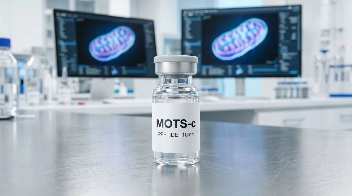
- MOTS-c is a 16-amino-acid mitochondrial-derived peptide encoded by a short open reading frame within the 12S rRNA gene of the mitochondrial genome.
- The originally proposed MOTS-c mechanism is indirect: inhibition of the folate cycle and tethered de novo purine biosynthesis causes AICAR to accumulate, which in turn activates AMPK.
- Skeletal muscle is the tissue where MOTS-c metabolic effects are most consistently reproduced, with rodent clamp studies attributing improved glucose handling to muscle clearance rather than hepatic suppression.
- MOTS-c and Humanin are both mitochondrial-derived peptides but differ in encoding gene, length, and mechanism: Humanin has a defined CNTFR/WSX-1/gp130 receptor complex, while MOTS-c has no established receptor.
- SS-31 (elamipretide) is a synthetic membrane-targeted tetrapeptide that acts on the mitochondrion, whereas MOTS-c is an endogenous signaling peptide that acts on the cell.
- The exercise mimetic label reflects overlap in a subset of metabolic pathway signatures, not equivalence to physical training.
- The m.1382A>C polymorphism (rs111033358) produces a reduced-activity K14Q MOTS-c variant associated in Japanese cohorts with type 2 diabetes risk in low-activity men and with muscle fiber composition and strength phenotypes.
- NCT07505745 is a registered Phase 2a randomized, quadruple-blind, placebo-controlled study of MOTS-c for insulin sensitivity in 120 adults with prediabetes, currently enrolling with no results posted.
- No human pharmacokinetic profile has been published for native MOTS-c; pharmacokinetic data from the CB4211 analog cannot be assigned to the unmodified peptide.
MOTS-c is a 16-amino-acid peptide encoded inside mitochondrial DNA rather than in the nucleus, and nearly everything known about it comes from cell and rodent work published since 2015. That gap between mechanistic depth and human evidence is where most MOTS-c content goes wrong. This article covers where the peptide actually comes from, what the AMPK evidence does and does not establish, how MOTS-c differs mechanistically from Humanin and SS-31, why the "exercise mimetic" label is an incomplete description, and what the first registered clinical trial of MOTS-c itself is testing.

MOTS-C
RESEARCH PEPTIDE
MOTS-C is a mitochondrial-derived peptide (MDP) made up of 16 amino acids and encoded within a short open reading frame of the mitochondrial genome, rather than nuclear DNA. Since its identification, it has become one of the more closely studied peptides in mitochondrial biology, largely because it appears to act as a signaling molecule between mitochondria and the rest of the cell. Helix Bio supplies MOTS-C as a lyophilized research peptide, manufactured to a purity standard of 99% or higher and tested in-house before every batch ships. It is sold exclusively for laboratory research use by qualified professionals — not for human or animal use, and not for human consumption.

SS-31
RESEARCH PEPTIDE
SS-31, also known as elamipretide, MTP-131, or Bendavia, is a synthetic mitochondria-targeted tetrapeptide studied extensively in laboratory research involving mitochondrial membranes, cardiolipin interactions, cellular bioenergetics, oxidative stress models, and mitochondrial protein interactions. SS-31 is particularly relevant to researchers investigating how small cationic-aromatic peptides interact with mitochondrial membranes and influence molecular processes associated with mitochondrial structure and function. Research has identified interactions between SS-31 and cardiolipin-containing membranes, as well as several mitochondrial proteins associated with energy metabolism. Helix Bio supplies research peptides for laboratory and scientific applications. The company's website states that its products are research and laboratory materials rather than products intended for human or veterinary use.
What Is MOTS-c Peptide?
MOTS-c is a mitochondrial-derived peptide composed of 16 amino acids, encoded by a short open reading frame within the 12S rRNA region of the mitochondrial genome. Its name is an acronym: mitochondrial open reading frame of the 12S rRNA type-c. It is studied as a signaling molecule that appears to communicate mitochondrial metabolic state to the rest of the cell, rather than as a structural or catalytic component of energy production.
The distinction matters. The mitochondrial genome was long described as encoding 13 proteins, 22 tRNAs, and 2 rRNAs, all of which serve oxidative phosphorylation or the machinery that builds it. MOTS-c belongs to a different category: a mitochondrial-derived peptide, or MDP, translated from a short open reading frame nested inside a gene that was assumed to be non-coding for proteins.
Attribute | Detail |
|---|---|
Full name | Mitochondrial open reading frame of the 12S rRNA type-c |
Class | Mitochondrial-derived peptide (MDP) |
Length | 16 amino acids |
Sequence | MRWQEMGYIFYPRKLR |
Genomic origin | Short ORF within MT-RNR1 (12S rRNA), mitochondrial genome |
Year first described | 2015 |
Principal research themes | Insulin sensitivity, skeletal muscle glucose metabolism, AMPK signaling, exercise physiology, aging |
Human evidence stage | Genetic and observational data; one registered interventional trial, no published results |
Where Does MOTS-c Come From?
The mitochondrial genome contains two ribosomal RNA genes: MT-RNR1, encoding the 12S rRNA, and MT-RNR2, encoding the 16S rRNA. Both turn out to harbor short open reading frames that produce small peptides. The 16S region encodes Humanin and a family of small humanin-like peptides. The 12S region encodes MOTS-c.
Two details about MOTS-c biogenesis are frequently reported incorrectly. First, MOTS-c is translated in the cytosol using the standard genetic code, not inside the mitochondrion using the mitochondrial code — which is why its open reading frame reads as a coherent peptide only when interpreted cytoplasmically. Second, MOTS-c is not a fragment of a larger mitochondrial protein that has been cleaved. It is the direct product of its own short reading frame.
That genomic address is also why a single nucleotide change in the 12S rRNA gene can alter both the ribosomal RNA and the peptide sequence at the same time. This overlap becomes important in the human genetic evidence discussed below.
Retrograde Mitochondrial Signaling
Retrograde signaling describes information flowing from mitochondria back toward the cytosol and nucleus, in the opposite direction to the more familiar anterograde control the nucleus exerts over mitochondria. Most retrograde signals studied before MDPs were metabolites or stress intermediates: calcium, reactive oxygen species, NAD+ and AMP ratios, and unfolded protein responses.
Mitochondrial-derived peptides added a different possibility — that the organelle encodes and releases discrete peptide messages of its own. In cell models, MOTS-c has been observed to move into the nucleus under metabolic stress and to associate with regulatory regions of stress-responsive nuclear genes. That observation is the strongest conceptual reason MOTS-c attracts attention: it suggests the mitochondrial genome participates in nuclear gene regulation rather than only responding to it.
This is mechanistic and largely in vitro evidence. It establishes that the interaction can occur in the systems tested. It does not establish how much of any whole-organism effect is attributable to nuclear translocation versus other actions.
MOTS-c and AMPK Activation
AMP-activated protein kinase (AMPK) is the cell's principal low-energy sensor. When the AMP-to-ATP ratio rises, AMPK activation shifts the cell away from anabolic, ATP-consuming processes and toward catabolic ATP-generating ones, including glucose uptake and fatty acid oxidation.
The originally proposed MOTS-c mechanism is indirect and worth stating precisely, because it is usually compressed into the phrase "MOTS-c activates AMPK" and loses its content in the process.
The folate cycle step most summaries skip
In the founding work, MOTS-c was reported to inhibit the folate cycle and the de novo purine biosynthesis pathway tethered to it. One-carbon units carried by folate derivatives are required to build purine rings. Interrupting that flux causes the intermediate AICAR to accumulate. AICAR is a recognized endogenous AMPK activator. On this model, MOTS-c does not bind AMPK — it changes one-carbon metabolism such that an AMPK-activating intermediate builds up.
That is a more interesting claim than "activates AMPK," and it is also more fragile. It predicts that MOTS-c effects should depend on folate cycle flux, purine synthesis rates, and cell type, and it means AMPK activation is a downstream readout rather than a direct target engagement.
Where AMPK is not the whole story
Later work identified additional interactions that do not reduce to the folate–AICAR–AMPK chain. Research in skeletal muscle has implicated casein kinase 2 (CK2), with MOTS-c-induced muscle glucose uptake blunted by CK2 inhibition or CK2 alpha knockdown. Nuclear translocation under stress and effects on inflammatory signaling have also been described in models where AMPK is not the sole explanatory pathway.
The honest summary is that AMPK-related signaling is the most consistently reported downstream feature of MOTS-c exposure across models, that at least two distinct routes to it have been proposed, and that no single receptor has been established for MOTS-c in the way one has for Humanin or for GLP-1 agonists.
Skeletal Muscle and Glucose Metabolism
Skeletal muscle is the tissue where MOTS-c metabolic effects have been most consistently reproduced. In rodent work, systemic MOTS-c administration improved glucose tolerance, and hyperinsulinemic-euglycemic clamp studies indicated the effect operated through increased skeletal muscle glucose clearance rather than through suppression of hepatic glucose production. Rodent studies have also reported protection against diet-induced insulin resistance and diet-induced obesity, and restoration of muscle insulin sensitivity in middle-aged animals.
Endogenous levels move with metabolic state. Prolonged fasting reduced MOTS-c in mouse skeletal muscle and plasma, which is consistent with a peptide whose expression tracks substrate availability and mitochondrial load.
Every finding in this section comes from cell culture or animal models. Rodent glucose-handling results do not transfer to humans by default, and none of these observations have been reproduced as clinical outcomes in a completed human trial of MOTS-c.
Is MOTS-c an Exercise Mimetic?
Partially, and only in a narrow technical sense. The exercise mimetic peptide label rests on two specific observations: exercise induces endogenous MOTS-c expression in human skeletal muscle and in circulation, and exogenous MOTS-c reproduces some exercise-associated transcriptional and metabolic signatures in animal and cell models. In one line of work, intermittent MOTS-c treatment begun late in life increased physical capacity in mice.
What the label does not mean is that MOTS-c reproduces the effects of physical training. Exercise simultaneously produces mechanical loading, cardiovascular adaptation, neural adaptation, bone loading, appetite and sleep effects, and a broad endocrine response involving dozens of signaling molecules. "Exercise mimetic" in the MDP literature is shorthand for overlap in a subset of metabolic pathway signatures. Treating it as a claim of equivalence is the single most common misreading of MOTS-c research.
A more defensible framing: MOTS-c behaves in these models like one component of the exercise response that can be studied in isolation. That is scientifically useful precisely because it is narrower than the popular framing, and it is why MOTS-c appears alongside growth-axis compounds in our muscle growth research comparison without belonging to the same mechanistic class.
MOTS-c and Aging Research
Three separate strands of evidence connect MOTS-c to aging, and they are not equally strong.
The first is descriptive: circulating MOTS-c has been reported to decline with age in human plasma, consistent with the broader pattern of declining MDP levels alongside mitochondrial dysfunction. This is an association, and declining levels do not establish that the decline is causal or that restoring levels would be beneficial.
The second is interventional but preclinical: late-life MOTS-c administration improved measures of physical capacity in aged mice. This is an animal healthspan result, not a demonstrated lifespan extension in humans.
The human genetic evidence
The third strand is the most interesting and least discussed. A polymorphism in the MOTS-c coding region, m.1382A>C (rs111033358), substitutes glutamine for lysine at position 14, producing a K14Q variant with reduced biological activity. The variant is essentially specific to Northeast Asian populations.
Research in Japanese cohorts has associated the C allele with increased type 2 diabetes prevalence in men, and notably found that association concentrated in men in the lowest tertile of physical activity — a gene-by-activity interaction. Separate work associated the same allele with a higher proportion of fast-twitch myosin heavy chain isoform, higher isokinetic peak torque in men, and a higher allele frequency in sprint and power athletes than in endurance athletes.
This is human evidence, and it is the closest thing to a causal human argument in the MOTS-c literature — a naturally occurring loss-of-function variant tracking with the metabolic and muscle phenotypes that the animal work predicts. It is also observational and population-specific, and one larger analysis found the polymorphism did not affect lifespan, which cuts against the simplest longevity interpretation.
MOTS-c vs Humanin
Both are mitochondrial-derived peptides. That shared class is where the similarity mostly ends. They come from different rRNA genes, have different lengths, act through different mechanisms, and are studied for different reasons.
Attribute | MOTS-c | Humanin |
|---|---|---|
Encoding gene | MT-RNR1 (12S rRNA) | MT-RNR2 (16S rRNA) |
Length | 16 amino acids | 24 amino acids |
Year first described | 2015 | 2001 |
Best-characterized mechanism | Folate cycle inhibition, AICAR accumulation, downstream AMPK signaling; CK2 interaction in muscle | Binds CNTFR/WSX-1/gp130 trimeric receptor driving JAK2/STAT3 signaling; binds BAX intracellularly to inhibit apoptosis |
Defined cell-surface receptor | Not established | Yes, characterized trimeric complex |
Primary research focus | Insulin sensitivity, skeletal muscle glucose metabolism, exercise physiology | Cytoprotection, anti-apoptotic signaling, neuroprotection |
Human interventional evidence | One registered Phase 2a trial, no published results | Essentially absent |
The clearest mechanistic contrast: Humanin has a defined receptor complex and a direct intracellular protein target, while MOTS-c is best described as a metabolic pathway modulator without an established receptor. Neither is "better." They answer different research questions, and a study designed around cell survival under stress is not interchangeable with one designed around muscle glucose disposal.
MOTS-c vs SS-31 (Elamipretide)
SS-31, also called elamipretide, is frequently grouped with MOTS-c as a "mitochondrial peptide," which obscures a fundamental difference. SS-31 is a synthetic aromatic-cationic tetrapeptide designed to concentrate at the inner mitochondrial membrane, where it associates with cardiolipin and influences membrane structure and electron transport chain organization. It is a targeted biophysical agent.
MOTS-c is an endogenous peptide encoded by mitochondrial DNA that acts as a signaling molecule outside the organelle. SS-31 acts on the mitochondrion; MOTS-c acts on the cell on behalf of the mitochondrion. SS-31 also carries a substantially longer clinical development record, including registered human trials in primary mitochondrial myopathy and other indications, which MOTS-c does not have. Researchers comparing the two should be comparing membrane-targeted structural intervention against signaling-pathway modulation, not two flavors of the same thing. Helix Bio supplies SS-31 as a separate research compound for exactly this reason.
MOTS-c vs GLP-1 Agonists and Semaglutide
This comparison is the one most likely to be abused, so it is worth being blunt: they are not in the same evidentiary category.
Semaglutide and related incretin-based compounds are receptor agonists with defined molecular targets, characterized human pharmacokinetics, completed Phase 3 programs, and regulatory approvals for specific indications. MOTS-c has no established receptor, no published human pharmacokinetic profile, and one ongoing early-phase trial with no results.
Dimension | MOTS-c | GLP-1 receptor agonists (e.g. semaglutide) |
|---|---|---|
Molecular target | No established receptor; pathway-level modulation | GLP-1 receptor, a defined class B GPCR |
Proposed mechanism | Folate cycle and AMPK-related metabolic signaling | Incretin receptor agonism affecting insulin secretion, gastric emptying, satiety signaling |
Primary tissue in research models | Skeletal muscle | Pancreatic islets, gut, central appetite circuits |
Human clinical stage | One registered Phase 2a study, no results | Completed Phase 3 programs, approved products |
Evidence level | Mechanistic, in vitro, animal, human genetic association | Human interventional, regulatory grade |
A researcher can reasonably study both in a metabolic model. What no one can currently support is any claim that MOTS-c substitutes for, outperforms, or is comparable to an incretin agonist in humans. The relevant receptor-level comparison of GLP-1 and GIP agonists sits in an entirely different evidence tier.
What Does the Current Human Research Actually Show?
Two items make up the human interventional record, and neither says what promotional content usually claims.
The CB4211 analog trials
CB4211 is a modified MOTS-c analog, not MOTS-c itself. CohBar ran a randomized, double-blind, placebo-controlled Phase 1a/1b program: a Phase 1a single- and multiple-ascending-dose stage in 65 healthy adults, followed by a Phase 1b stage giving 25 mg subcutaneously once daily for four weeks to 20 subjects with obesity and nonalcoholic fatty liver disease. The study met its primary safety and tolerability endpoint with no serious adverse events. Exploratory pharmacodynamic endpoints showed statistically significant reductions in ALT and AST versus placebo, a significant decrease in glucose, and a trend toward lower body weight.
Three qualifications matter more than the headline. These were exploratory endpoints in 20 subjects over four weeks, not efficacy endpoints in a powered trial. The compound was an analog with different properties from native MOTS-c. And the program's earlier stage had been temporarily suspended to address persistent injection site reactions.
NCT07505745
The first registered interventional trial of MOTS-c itself is NCT07505745, listed as MOTS-MET, sponsored by Hudson Biotech under protocol identifier EX-MOTS-2A-001.
Field | Registered detail |
|---|---|
Phase | 2a |
Design | Randomized, parallel assignment, quadruple-blind, placebo-controlled |
Population | 120 estimated participants, ages 18 to 65, BMI 27.0 to 40.0 kg/m2, documented prediabetes |
Intervention | Investigational MOTS-c by subcutaneous injection, or matching placebo, 1:1, both with standardized lifestyle counseling |
Duration | Up to 4-week screening, 12-week double-blind treatment, 4-week safety follow-up |
Efficacy assessment | 75 g OGTT-derived insulin sensitivity index, plus HbA1c, fasting glucose, lipids, body weight, waist circumference |
Status | Enrolling; no results posted |
A trial being registered and enrolling establishes that a hypothesis is being tested under controlled conditions. It does not establish that the hypothesis is correct. Until results are posted and peer-reviewed, NCT07505745 is evidence of clinical investigation underway, not evidence of clinical efficacy.
The registry entry itself is transparent about this framing: the rationale section describes AMPK activation as a proposed downstream mechanism and skeletal muscle as a key target tissue reported in preclinical studies, and states the trial is designed to test the hypothesis in humans.
What MOTS-c Half-Life Research Does and Does Not Establish
There is no established human half-life for MOTS-c, and any specific number presented as settled should be treated skeptically until the source is checked.
What exists is fragmentary and heterogeneous. Preclinical work has used varied routes, doses, and administration schedules — intraperitoneal in some rodent studies, intermittent dosing three times weekly in the healthspan work — which implies different effective durations of action but does not resolve into a single pharmacokinetic parameter. Pharmacokinetics were measured as a secondary endpoint in the CB4211 program, but CB4211 is an analog engineered for different properties, so its profile cannot be assigned to native MOTS-c. Short unmodified peptides are generally subject to rapid proteolytic clearance, which is a reasonable prior expectation and not a measurement.
The practical consequence for research design: half-life should be treated as an open variable to be determined in the specific model and assay being used, not as a published constant to be looked up.
Evidence Map
Research area | Model | Principal finding | Evidence level | Key limitation |
|---|---|---|---|---|
Genomic origin | Molecular biology | 16-aa peptide from short ORF in 12S rRNA gene | Established mechanistic | None material; well replicated |
Nuclear translocation | Cell culture | MOTS-c moves to nucleus under metabolic stress and associates with stress-response gene regulation | In vitro | Contribution to whole-organism effects unquantified |
Folate cycle and AMPK | Cell culture and rodent | Folate cycle inhibition, AICAR accumulation, downstream AMPK signaling | Mechanistic and in vitro | Indirect mechanism; not the only reported pathway |
Muscle glucose disposal | Rodent, clamp studies | Improved glucose clearance via skeletal muscle rather than hepatic suppression | Animal interventional | Species and model dependent |
Exercise induction | Human skeletal muscle and plasma | Exercise increases endogenous MOTS-c expression and circulating levels | Human observational | Correlation with exercise, not evidence that exogenous peptide reproduces exercise |
Age-related decline | Human plasma | Circulating MOTS-c declines with age | Human observational | Association only |
K14Q variant | Human genetic cohorts | Reduced-activity variant associated with T2D risk in low-activity men, and with muscle fiber and strength phenotypes | Human genetic association | Population-specific; no lifespan effect in larger analysis |
CB4211 analog | Human Phase 1a/1b | Safety endpoint met; exploratory ALT, AST, glucose changes vs placebo | Human interventional, early phase | Analog not MOTS-c; 20 subjects; exploratory endpoints |
MOTS-c clinical | Human Phase 2a, NCT07505745 | Insulin sensitivity in prediabetes under investigation | Clinical development | Enrolling; no results |
Research Material Quality, Storage and Documentation
For laboratory work, the questions that determine whether a MOTS-c experiment is interpretable are identity, purity, and lot traceability. A 16-residue peptide is short enough that synthesis-related impurities — deletion sequences, incomplete deprotection, residual counter-ions — can materially affect an assay without being visually apparent in a vial of white powder.
Two complementary analyses address this. High-performance liquid chromatography quantifies purity as a percentage of total peptide-related material. Mass spectrometry confirms that the observed molecular weight matches the intended sequence, which HPLC alone cannot do. A purity figure without an identity confirmation is an incomplete document, and both should be tied to the specific lot number printed on the vial.
Helix Bio supplies MOTS-C as a lyophilized research peptide, manufactured to a purity target of 99% or higher, verified by HPLC and mass spectrometry, with a batch-specific Certificate of Analysis available for each lot. The current product page specifies storage of lyophilized material at -20°C or colder protected from light, reconstitution with appropriate bacteriostatic or sterile water using standard laboratory technique, storage of reconstituted material at 2-8°C for the timeframe indicated on the batch documentation, and avoidance of repeated freeze-thaw cycles.
Solvent volume and post-reconstitution stability windows are lot-specific. Follow the current batch documentation and Certificate of Analysis for the vial in hand rather than a generic figure from a secondary source. Our [guide to reading a Certificate of Analysis](/how-to-read-peptide-certificate-of-analysis) walks through the fields that matter, and the [reconstitution and storage guide](/peptide-reconstitution-guide-bacteriostatic-water-storage-stability) covers general laboratory handling practice.
MOTS-c is not approved by the FDA for human use in any form, and it is prohibited in sport under anti-doping rules. Materials sold on a research-use-only basis are not evaluated for human safety or efficacy, a point covered in more depth in our overview of research peptide legality and RUO compliance.
Limitations and Open Questions
Several gaps in the MOTS-c literature are worth stating explicitly, because they define what a well-designed study can currently contribute.
- No receptor has been established. Without a defined binding partner at the cell surface, dose-response relationships, tissue selectivity, and off-target behavior are difficult to model.
- Mechanism is plural, not singular. Folate cycle inhibition, CK2 interaction in muscle, and nuclear translocation have each been reported; their relative contributions in any given tissue are unresolved.
- Human pharmacokinetics are absent for the native peptide. Everything published on PK concerns animals or an engineered analog.
- Sex differences are underexplored. Several reviews note that available evidence hints at differing responses between males and females, with dosing implications that have not been addressed.
- Endogenous versus exogenous effects may diverge. Exercise-induced endogenous MOTS-c and administered synthetic MOTS-c reach tissues by different routes and at different concentrations, and evidence for one does not automatically support the other.
- Assay heterogeneity limits comparison. Reported circulating MOTS-c concentrations vary considerably across studies using different immunoassays, which complicates cross-study synthesis of the age-decline literature.
What the field has, credibly, is a well-characterized genomic origin, a reproducible metabolic phenotype in rodents, a plausible and partially specified mechanism, and one line of human genetic evidence consistent with the preclinical model. What it does not have is a completed human interventional result. NCT07505745 is the first study positioned to change that, and until it reports, the accurate description of MOTS-c is a mechanistically interesting mitochondrial-derived peptide at an early stage of clinical investigation.
Frequently Asked Questions
MOTS-c is a 16-amino-acid mitochondrial-derived peptide encoded by a short open reading frame within the 12S rRNA region of mitochondrial DNA. Its name stands for mitochondrial open reading frame of the 12S rRNA type-c. It is studied in research models as a signaling molecule that communicates mitochondrial metabolic state to the rest of the cell.
The best-characterized proposed mechanism is indirect: MOTS-c inhibits the folate cycle and the de novo purine biosynthesis pathway tethered to it, causing the AMPK-activating intermediate AICAR to accumulate. Separate research in skeletal muscle has implicated casein kinase 2, and cell studies describe nuclear translocation under metabolic stress. No cell-surface receptor for MOTS-c has been established.
Both are mitochondrial-derived peptides, but MOTS-c is 16 amino acids encoded in the 12S rRNA gene while Humanin is 24 amino acids encoded in the 16S rRNA gene. Humanin signals through a defined CNTFR/WSX-1/gp130 trimeric receptor and binds BAX intracellularly to inhibit apoptosis; MOTS-c is studied as a metabolic pathway modulator without an established receptor. Humanin research centers on cytoprotection, MOTS-c research on insulin sensitivity and muscle glucose metabolism.
A mitochondrial-derived peptide is a small peptide translated from a short open reading frame located inside a mitochondrial gene that was previously assumed not to encode proteins. Known MDPs arise from the 12S and 16S rRNA genes and include MOTS-c, Humanin, and the small humanin-like peptides. They are studied as retrograde signals, meaning information flowing from the mitochondrion outward rather than nuclear control flowing in.
Only in a narrow technical sense. Exercise induces endogenous MOTS-c expression in human skeletal muscle and circulation, and exogenous MOTS-c reproduces some exercise-associated metabolic signatures in animal and cell models. It does not reproduce the mechanical, cardiovascular, neural, and broader endocrine components of training, so the label describes pathway overlap rather than equivalence.
The connection is mechanistic and preclinical. In cell and rodent models, MOTS-c exposure inhibits folate-dependent one-carbon flux, AICAR accumulates, and AMPK signaling increases downstream. AMPK activation in this model is a readout rather than a direct binding event, and later work has identified additional pathways that AMPK alone does not explain.
Rodent studies report that MOTS-c administration improves glucose tolerance and protects against diet-induced and age-related insulin resistance, with hyperinsulinemic-euglycemic clamp data attributing the effect to skeletal muscle glucose clearance. In humans, the evidence is genetic and observational rather than interventional, and no completed clinical trial has demonstrated an insulin sensitivity outcome for MOTS-c.
NCT07505745 is a registered Phase 2a randomized, quadruple-blind, placebo-controlled study evaluating whether 12 weeks of subcutaneous MOTS-c improves insulin sensitivity versus placebo in 120 adults aged 18 to 65 with prediabetes and a BMI of 27.0 to 40.0. It is listed as enrolling with no results posted. Registration and enrollment establish that the hypothesis is being tested, not that it has been confirmed.
GLP-1 receptor agonists such as semaglutide act on a defined class B GPCR with characterized human pharmacokinetics and completed Phase 3 programs. MOTS-c has no established receptor, no published human pharmacokinetic profile, and one ongoing early-phase trial. They occupy different evidence tiers, and no current data supports treating MOTS-c as comparable to an incretin agonist in humans.
SS-31 is a synthetic aromatic-cationic tetrapeptide designed to concentrate at the inner mitochondrial membrane and associate with cardiolipin, so it acts structurally on the organelle itself. MOTS-c is an endogenous mitochondrially encoded peptide that acts as a signal outside the organelle. SS-31 also carries a substantially longer clinical development record than MOTS-c.
No human half-life has been published for native MOTS-c. Preclinical studies have used varied routes, doses, and dosing intervals without resolving into a single pharmacokinetic parameter, and the pharmacokinetic data collected in the CB4211 program describe an engineered analog rather than the unmodified peptide. Researchers should determine effective duration within their own model rather than citing a fixed figure.
Per the current Helix Bio product documentation, lyophilized MOTS-C is stored at -20°C or colder protected from light, reconstituted with appropriate bacteriostatic or sterile water using standard laboratory technique, and stored at 2-8°C after reconstitution for the timeframe indicated on the batch documentation, avoiding repeated freeze-thaw cycles. Solvent volume and stability windows are lot-specific, so the Certificate of Analysis for the vial in hand takes precedence over generic figures.
- 01Reynolds JC, Lai RW, Woodhead JST, et al. MOTS-c is an exercise-induced mitochondrial-encoded regulator of age-dependent physical decline and muscle homeostasis. Nature Communications. 2021;12:470.
- 02Kim KH, Son JM, Benayoun BA, Lee C. MOTS-c: A novel mitochondrial-derived peptide regulating muscle and fat metabolism. Free Radical Biology and Medicine. 2016.
- 03Zheng Y, Wei Z, Wang T. MOTS-c: A promising mitochondrial-derived peptide for therapeutic exploitation. Frontiers in Endocrinology. 2023;14:1120533.
- 04Zempo H, Kim SJ, Fuku N, et al. A pro-diabetogenic mtDNA polymorphism in the mitochondrial-derived peptide, MOTS-c. Aging. 2021;13(2):1692-1717.
- 05Kumagai H, Natsume T, Kim SJ, et al. The MOTS-c K14Q polymorphism in the mtDNA is associated with muscle fiber composition and muscular performance. Biochimica et Biophysica Acta - General Subjects. 2022;1866(1):130048.
- 06Merry TL, Chan A, Woodhead JST, et al. Mitochondrial-derived peptides in aging and age-related disease. Review of MDP biology, 12S and 16S rRNA origins, and MOTS-c function.
- 07ClinicalTrials.gov. MOTS-c for Improving Insulin Sensitivity in Adults With Prediabetes and Overweight/Obesity (MOTS-MET), NCT07505745. Sponsor: Hudson Biotech. Phase 2a, enrolling.
- 08CohBar, Inc. CohBar Announces Positive Topline Results from the Phase 1a/1b Study of CB4211 Under Development for NASH and Obesity. Press release, August 10, 2021.

Helix Bio Chem Team
Research & Product Team
Our in-house team tracks published peptide research and translates it into clear, source-cited summaries for the research community.
Reviewed by in-house research chemists
support@helixbiochem.com