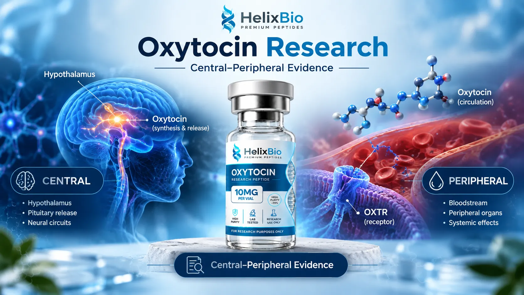
Oxytocin operates as two separately regulated pools, central and peripheral, that are divided by the blood-brain barrier and do not reliably correlate.
Magnocellular neurons of the paraventricular and supraoptic nuclei supply the peripheral pool via the posterior pituitary, while a separate central projection and dendritic release supply the brain.
CD38 generates cyclic ADP-ribose and is required for normal oxytocin secretion; CD38 knockout mice show reduced plasma and cerebrospinal fluid oxytocin.
The oxytocin receptor is a class A GPCR signalling principally through Gq/11, phospholipase C, IP3 and intracellular calcium, with Gi/o coupling also reported.
Plasma oxytocin measured with and without solid-phase extraction differs by roughly an order of magnitude and the two measurements do not correlate, so studies using different methods should not be pooled.
Intranasal oxytocin produces a measurable but delayed cerebrospinal fluid rise, peaking around 75 minutes against a plasma peak near 15 minutes, with no correlation between compartments.
A widely cited critical analysis estimates that roughly 0.005% of an intranasally administered oxytocin dose reaches cerebrospinal fluid.
A 24-week multi-site randomised trial of intranasal oxytocin in autism spectrum disorder found no significant difference from placebo on its primary social outcome.
Strong clinical evidence for oxytocin belongs to an approved injectable obstetric product standardised in units of biological activity, not to research material.
Oxytocin never entered the 503A compounding bulk substances framework because it is an approved drug's active ingredient with a pharmacopoeial monograph.
Oxytocin has a longer research record than almost any other peptide and one of the least settled evidence bases in contemporary neuroscience. The molecular facts are not in dispute: the sequence, the receptor and the primary signalling cascade are all well characterised. What remains contested sits downstream of that chemistry — how much of an administered dose reaches the brain, whether circulating oxytocin reports on central oxytocin at all, and how much of the behavioural literature survives replication. This article covers the evidence layer rather than the entity layer. Molecular identity, analytical specifications and receptor identifiers are set out on the Oxytocin product page; what follows is what the published research does and does not establish.
Featured In This Article
Oxytocin
RESEARCH PEPTIDE
Highly purified synthetic peptide prepared for rigorous laboratory research.
$71.99
Two oxytocin systems that share one name
The single most common error in secondary writing on this topic is treating oxytocin as one circulating quantity. It is not. Oxytocin operates as two functionally distinct pools that are separated by the blood-brain barrier and are not reliably correlated with one another.
The peripheral pool
Magnocellular neurons of the hypothalamic paraventricular and supraoptic nuclei project axons to the posterior pituitary, where oxytocin is released into systemic circulation as a classical neurohormone. This pool acts on oxytocin receptors in myometrium and mammary myoepithelium, and it is the pool measured by any blood draw. Circulating oxytocin is cleared rapidly, with a plasma half-life measured in minutes.
The central pool
A separate population of oxytocin neurons projects within the brain itself, releasing peptide onto forebrain and limbic targets. Oxytocin neurons also release peptide from their dendrites and soma inside the hypothalamus, a mode of release that is regulated independently of axonal secretion from the pituitary. The practical consequence is that central release and pituitary release can be dissociated: one can occur without the other. Any experimental design that infers central oxytocin activity from a peripheral measurement is making an assumption the physiology does not support.
Synthesis, processing and release
Oxytocin is translated as a larger precursor, prepro-oxytocin, which is processed to yield the mature nonapeptide together with its carrier protein neurophysin I. The two are packaged in the same neurosecretory vesicles and travel together down the axon, which is why neurophysin I appears as an analyte in some research contexts and as a potential co-eluting species in others.
CD38 and the release machinery
Release is not simply a function of peptide availability. CD38, an ectoenzyme that generates cyclic ADP-ribose, mobilises intracellular calcium in oxytocin neurons and is required for normal secretion. CD38 knockout mice show reduced plasma and cerebrospinal fluid oxytocin alongside altered social behaviour, and the phenotype is rescued by CD38 re-expression or oxytocin administration. This is a useful reminder that a research model probing oxytocin biology can target the release machinery rather than the receptor, and that the two produce different readouts.
When designing around endogenous release, note that dendritic and axonal release are separately regulated. A manipulation that raises plasma oxytocin does not establish that central release changed, and the reverse is equally true.
What the receptor cascade establishes
The oxytocin receptor is a class A G protein-coupled receptor and, so far, the only identified receptor for the peptide. Its principal transduction route runs through Gq/11 to phospholipase C, generating inositol trisphosphate and diacylglycerol, mobilising intracellular calcium and activating protein kinase C. Coupling to Gi/o has also been reported, and pathway selection appears to vary by tissue and by receptor environment rather than being fixed.
That cascade is well established at the level of cells and tissues. It does not by itself predict a behavioural outcome in an intact animal, and no amount of receptor-level detail closes the gap between a calcium transient in a myometrial cell and a social phenotype in a mouse. Treating mechanism as evidence of effect is the most common inferential error in this literature.
Receptor selectivity is not absolute
Oxytocin and arginine vasopressin differ at two of nine positions, and their receptors are close relatives within the same GPCR family. Oxytocin retains appreciable affinity at vasopressin receptors, particularly V1a, and vasopressin retains affinity at the oxytocin receptor. At the concentrations reached in a binding assay this is manageable and quantifiable. In an intact animal given a supraphysiological dose it is not trivially separable, because an observed response may be mediated at a vasopressin receptor rather than at the oxytocin receptor.
The consequence for study design is specific: an effect observed after administering oxytocin is not attributable to the oxytocin receptor unless the design includes a selective antagonist, a receptor knockout, or another means of assigning the response to a receptor. A large share of the in vivo literature does not include one, which is a separate limitation from the delivery and measurement problems described below and compounds with them.
The measurement problem
Before any oxytocin result can be interpreted, the assay behind it has to be interrogated. This is not a minor methodological footnote — it is the reason a substantial fraction of the published human literature is difficult to compare.
Extracted versus unextracted immunoassay
Plasma oxytocin measured by immunoassay after solid-phase extraction and plasma oxytocin measured by the same immunoassay without extraction return values that differ by roughly an order of magnitude, and the two measurements do not correlate with each other within the same samples. Unextracted assays appear to detect immunoreactive material other than free oxytocin. A study reporting a plasma oxytocin association is therefore reporting something different depending on whether the sample was extracted, and studies using the two approaches should not be pooled.
Why plasma is a poor proxy for the brain
Because the two pools are separately regulated and the barrier between them is real, plasma concentration cannot stand in for central concentration. Cerebrospinal fluid sampling is closer to the target compartment but is invasive, sparsely timed in most protocols, and still an imperfect proxy for extracellular concentration at a receptor.
A reported plasma oxytocin value is uninterpretable without three pieces of information: the assay platform, whether the sample was extracted before assay, and the sampling time relative to any intervention. Research summaries that omit all three should be treated as unciteable.
Intranasal administration and the CSF question
Intranasal delivery is the route used in most human behavioural studies, and it is where the evidence is thinnest relative to the confidence with which it is usually described. The general question of how any peptide reaches the central nervous system is covered separately in how peptides reach the brain; what follows is specific to oxytocin.
Controlled human work with combined blood and cerebrospinal fluid sampling has shown that intranasal oxytocin does raise CSF oxytocin above placebo. The kinetics are informative: plasma concentrations peak early, within roughly fifteen minutes, while the CSF rise is delayed and is typically detected around seventy-five minutes. The two compartments do not track each other, which argues against the CSF signal being simple passive diffusion from blood — but sample sizes in these studies are small and CSF sampling points are few.
Compartment
Approximate time to peak
Correlates with the other compartment?
Plasma
~15 minutes
No
Cerebrospinal fluid
~75 minutes
No
The magnitude is the part most often dropped. A widely cited critical analysis estimates that on the order of 0.005% of an intranasally administered dose reaches cerebrospinal fluid. That figure reframes the question entirely. "Intranasal oxytocin reaches the brain" is defensible as a statement about detectability. It is not defensible as a statement about achieving a pharmacologically meaningful central concentration, and the two claims are routinely conflated. Reported nasal bioavailability is also low and varies substantially between individuals, so nominal dose is a weak predictor of exposure in any given subject.
A peripheral route to a central effect is not excluded
Even where a behavioural change is observed after intranasal administration, direct nose-to-brain transport is only one candidate explanation. Intranasal dosing reliably raises circulating oxytocin, and peripheral oxytocin can influence central state indirectly through autonomic and vagal afferent signalling, through cardiovascular and autonomic changes, or simply through the subjective experience of receiving a nasal spray. Distinguishing these possibilities requires a design that separates them, such as a matched intravenous comparator arm. Studies that administer intranasally and attribute the result to central receptor engagement are inferring the mechanism rather than testing it.
The human trial record
Early human behavioural findings — most famously an increase in trust in an economic game — generated an enormous secondary literature. Critical reviews of that body of work identified small sample sizes, flexible analysis choices and publication bias, and subsequent registered replications have generally failed to reproduce the original trust effect.
The largest and most decisive test to date was conducted in autism spectrum disorder. A multi-site randomised trial administered intranasal oxytocin or placebo daily for twenty-four weeks in children and adolescents and found no significant difference from placebo on its primary social withdrawal outcome. The trial was well powered and adequately controlled, which makes a null result substantially more informative than the many small positive studies that preceded it.
Evidence type
Strength for oxytocin
What it supports
Obstetric randomised evidence
Strong
Uterine contraction and postpartum haemorrhage control with the approved pharmaceutical product
Species-specific receptor distribution and social behaviour effects
Human pharmacokinetics
Moderate
Detectable CSF rise after intranasal dosing, with low magnitude
Human behavioural trials
Weak and inconsistent
No reliable prosocial effect at the level of a powered trial
Comparative animal work remains genuinely informative but does not transfer cleanly. Oxytocin receptor distribution differs markedly between species, including between closely related vole species with different social structures, so a behavioural result obtained in one rodent species is not a prediction about another, let alone about humans. The same caution applies to other hypothalamic neuropeptides with rich mechanistic literatures and thin translational records, a pattern also visible in kisspeptin research and VIP research.
Obstetric evidence belongs to a different product
The strong clinical evidence for oxytocin concerns an approved injectable pharmaceutical administered intravenously or intramuscularly under clinical supervision for labour induction, labour augmentation and control of postpartum bleeding. That evidence attaches to a specific manufactured product with a specific formulation, specification and route.
Two details are worth knowing because they invert a common assumption. The approved product is standardised in units of biological activity rather than by peptide mass, and its labelled specification permits a substantial fraction of total impurities. An approved medicine is therefore not automatically the analytically purer article, and its specification cannot be used as a benchmark for a research reference material — the two are characterised on different bases entirely.
Where oxytocin sits in the regulatory picture
Oxytocin occupies an unusual regulatory position among peptides discussed in research contexts. It is the active ingredient of an approved drug and has a pharmacopoeial monograph, which means it never entered the compounding bulk substances framework that governs unapproved peptides such as BPC-157 or semax. There is no advisory committee vote to cite, no category placement and no nomination history, because the substance was never a candidate for that pathway.
Separately, no intranasal oxytocin product is approved for any behavioural, psychiatric or wellness indication in the United States. Research material supplied for laboratory use is a third category again: it shares molecular identity with the approved active and none of its regulatory status, formulation or intended use.
Approval, indications and label specifications attaching to a pharmaceutical oxytocin product do not transfer to research material. Research-use-only oxytocin is not intended for human or veterinary use, ingestion, injection, nasal administration or any other route.
What this means for selecting research material
For laboratory work, the evidence picture translates into a short set of practical checks. Confirm which oxytocin pool an experimental readout is actually reporting on. Specify the assay and the extraction step in the protocol before collecting samples, not afterwards. Treat route as a variable with its own evidence requirements rather than an implementation detail.
On the material itself, oxytocin's degradation chemistry means an area-percent purity figure is only meaningful if the chromatographic method resolves the species that matter — oligosulfide variants arising from the disulfide bridge and covalently linked dimers. The general approach to interrogating those numbers is covered in the guide to reading a certificate of analysis, and the compound-specific figures and identifiers sit on the Oxytocin page.
The honest summary of the oxytocin literature is that a well-characterised molecule with an unambiguous receptor has produced a behavioural evidence base that has not held up under powered testing. That is a useful thing to know before designing a study around it, and it is the part most secondary sources leave out.
Got Questions?
Frequently Asked Questions
Research establishes oxytocin's molecular identity, its single known receptor and its Gq/11-coupled signalling cascade with high confidence. It establishes obstetric pharmacology for the approved injectable product. It does not currently establish reliable behavioural effects of intranasal administration in powered human trials.
The two pools are released by different neuronal populations and are separated by the blood-brain barrier. Dendritic and central release can occur independently of pituitary release into blood, so a change in one compartment does not imply a change in the other.
No. Plasma and central oxytocin are separately regulated, and human studies sampling both compartments after intranasal administration have found no correlation between them. Inferring central activity from a blood measurement is not supported by the physiology.
Plasma assayed with and without solid-phase extraction yields values differing by roughly an order of magnitude, and the two do not correlate within the same samples. Unextracted assays appear to detect immunoreactive material besides free oxytocin, so the extraction step must be reported for a result to be interpretable.
Controlled human studies show a measurable rise in cerebrospinal fluid oxytocin after intranasal administration, so it is detectable centrally. The quantity is very small — one widely cited estimate places it near 0.005% of the administered dose — so detectability should not be read as evidence of a pharmacologically meaningful central concentration.
In human studies with combined sampling, plasma concentrations peak at roughly fifteen minutes while the cerebrospinal fluid rise is detected around seventy-five minutes. The absence of correlation between compartments argues against simple passive diffusion from blood, though sample sizes in these studies are small.
A multi-site randomised placebo-controlled trial administering intranasal oxytocin daily for twenty-four weeks in children and adolescents with autism spectrum disorder found no significant difference from placebo on its primary social withdrawal outcome. Its size and design make the null result more informative than earlier small positive studies.
Critical reviews identified small samples, analytic flexibility and publication bias in that literature, and subsequent registered replication attempts have generally failed to reproduce the original effect. The finding should be treated as unreplicated rather than established.
Only with caution. Oxytocin receptor distribution differs markedly between species, including between closely related vole species with different social structures, so a behavioural result in one species is not a prediction for another.
No. Pharmaceutical oxytocin is an approved injectable product standardised in units of biological activity and manufactured to a defined specification for obstetric indications. Research-use-only material shares molecular identity and none of that regulatory status, formulation or intended use.
Outside it. Because oxytocin is the active ingredient of an approved drug and carries a pharmacopoeial monograph, it never entered the 503A bulk substances categories that apply to unapproved peptides, so there is no category placement or advisory committee vote associated with it.
That the chromatographic method resolves the degradation species this molecule actually produces. Oxytocin's disulfide bridge gives rise to oligosulfide variants and covalently linked dimers, and a method that does not separate them will report an area-percent figure that overstates intact peptide content.