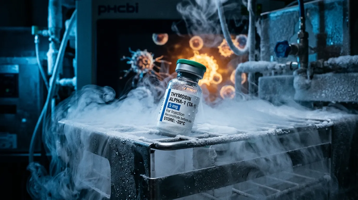
- Thymosin alpha-1 is a 28-amino-acid, N-terminally acetylated peptide corresponding to the first 28 residues of prothymosin alpha, with a molecular weight of approximately 3,108 Da.
- Thymosin alpha-1, Tα1, TA-1, thymalfasin and Zadaxin all refer to the same peptide sequence in different regulatory and commercial contexts.
- TA-1 is described as an immune modulator rather than an immune stimulant because its reported effects are context-dependent, amplifying responses to viral stimuli while dampening responses to bacterial stimuli in the same system.
- The proposed mechanism centres on Toll-like receptor signaling in dendritic cells, with TLR9/MyD88/IRF7 and TLR2/NF-κB pathways the best supported in the literature.
- Thymosin alpha-1 is not FDA-approved in the United States for any indication, despite thymalfasin being approved as a prescription medicine in more than 35 other countries.
- Thymosin alpha-1 was placed in FDA Category 2 in September 2023, not Category 1, and does not appear on the 503A Bulks List as of August 2026.
- The TESTS phase 3 trial (BMJ, 2025) found no reduction in 28-day sepsis mortality with thymosin alpha-1 in 1,106 patients, contradicting earlier meta-analyses of smaller trials.
- Thymosin alpha-1 and TB-500 are unrelated peptides: TA-1 derives from prothymosin alpha and is studied for immune modulation, while TB-500 is a thymosin beta-4 fragment studied for tissue repair.
Thymosin alpha-1 sits in an unusual position in peptide science. It is one of the most clinically studied peptides in existence, approved as a prescription medicine in more than 35 countries, and simultaneously not approved by the FDA for any indication in the United States. It has a plausible, well-mapped mechanism involving Toll-like receptors and dendritic cells, and it has also produced a large, well-powered randomized trial that found no benefit on its primary endpoint. Both of those things are true at once, and holding them together is what serious thymosin alpha-1 research actually requires.
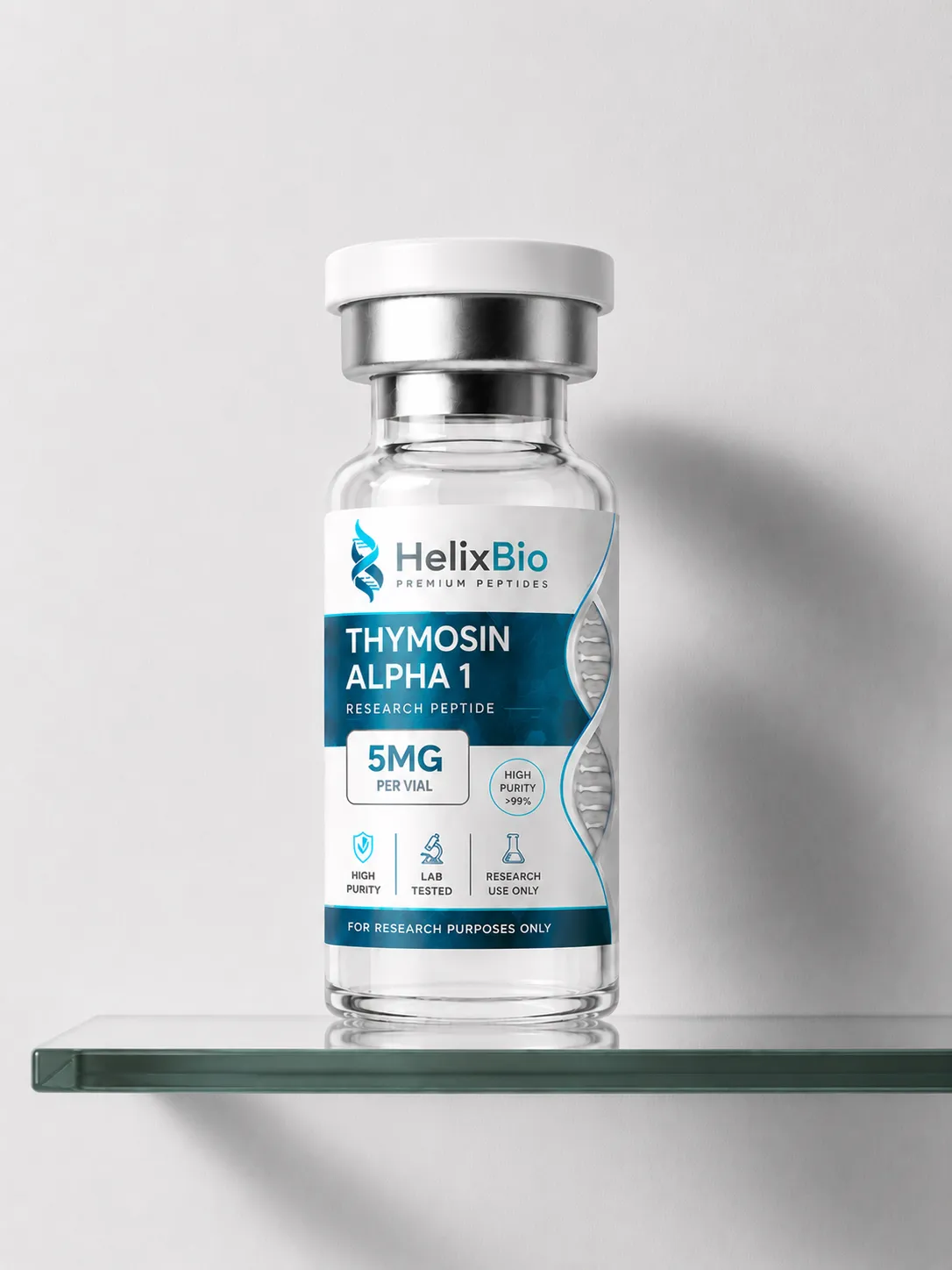
Thymosin Alpha-1
RESEARCH PEPTIDE
Thymosin Alpha-1 (Ta1) is a 28-amino-acid thymic peptide supplied by Helix Bio for laboratory and academic research use only. It is not manufactured, labeled, or sold for human consumption, clinical use, or self-administration of any kind, and it is not FDA-approved for any indication in the United States. Researchers working on innate and adaptive immune function, T-cell activity, cytokine regulation, or dendritic cell research use Thymosin Alpha-1 as a reference peptide because it's one of the more extensively studied thymic peptides in the immunology literature. Helix Bio's Thymosin Alpha-1 ships as a lyophilized powder, is verified through HPLC and mass spectrometry, and comes with a certificate of analysis for every batch. If you're comparing Thymosin Alpha-1 suppliers for a research order, the sections below cover specifications, storage guidance, and the published research context most researchers want before ordering.
This article covers what the TA-1 peptide is, how the proposed mechanism works at the level of T-cells, natural killer cells and TLR signaling, what the hepatitis B, hepatitis C and sepsis literature shows, how TA-1 differs from TB-500, and where U.S. regulatory status actually stands as of 2026. Where the phrase "viral protocols" appears, it refers to published clinical study designs and laboratory research protocols — not to instructions for administration. Helix Bio supplies research materials for laboratory use only.
What Is Thymosin Alpha-1?
Thymosin alpha-1 (Tα1, TA-1) is a 28-amino-acid peptide with an acetylated N-terminus and a molecular weight of approximately 3,108 Da. It was the first peptide isolated from thymic tissue, identified by Allen Goldstein and colleagues in 1977 during the fractionation of a bovine thymus extract known as thymosin fraction 5.
Structurally, TA-1 corresponds to the N-terminally acetylated first 28 residues of prothymosin alpha, a chromatin-associated precursor protein. Circulating TA-1 is generated by proteolytic cleavage of that precursor. The peptide is strongly acidic, contains no cysteine residues and therefore no disulfide bonds, and behaves as an intrinsically disordered peptide in aqueous solution at neutral pH — a structural detail that matters when researchers interpret binding studies, because TA-1 does not present a rigid, pre-formed binding surface the way a folded ligand does.
The name that appears in clinical literature is thymalfasin, the International Nonproprietary Name for the synthetic version of the same 28-residue sequence.
Attribute | Detail |
|---|---|
Common names | Thymosin alpha-1, Tα1, TA-1, thymalfasin |
Brand name (non-U.S.) | Zadaxin |
Length | 28 amino acids |
Modification | N-terminal acetylation |
Molecular weight | ~3,108 Da |
Origin | N-terminal fragment of prothymosin alpha |
First isolated | 1977, from thymosin fraction 5 |
Main research areas | Immune modulation, chronic viral infection, sepsis, oncology adjunct |
U.S. regulatory status | Not FDA-approved for any indication |
Tα1, Thymalfasin and Zadaxin: One Molecule, Three Names
Much of the confusion in search results around this compound is terminological rather than scientific. Thymosin alpha-1 is the endogenous peptide. Thymalfasin is the nonproprietary name of the synthetic pharmaceutical form of that same sequence. Zadaxin is the brand name under which thymalfasin is marketed internationally.
They refer to the same 28-residue molecule. What differs is the regulatory and manufacturing context: thymalfasin sold as Zadaxin is a licensed prescription pharmaceutical in the jurisdictions that approved it, manufactured to pharmaceutical standards and dispensed against a prescription. A thymalfasin peptide supplied as a research material is not that product, is not a drug, and carries no approved indication anywhere.
The practical consequence is that an approval in one country does not transfer. Zadaxin's approval in more than 35 countries, primarily for chronic hepatitis B and as an immune adjunct in oncology, establishes nothing about U.S. regulatory status.
Why TA-1 Is Called an Immune Modulator, Not an Immune Stimulant
The phrase TA-1 immune modulation is used loosely in vendor content, usually as a synonym for "boosts immunity." The published mechanism does not support that reading, and the distinction is the single most useful thing to understand about this peptide.
Immune modulation means the direction of the effect depends on the state of the system being acted on. The clearest demonstration comes from work by Giacomini and colleagues, who found that Tα1 amplified responses to viral-pattern stimuli while dampening responses to bacterial-pattern stimuli in the same experimental system. A compound that only stimulated would not do that. This context dependence is also why TA-1 has been described as a pleiotropic immune modifier — pleiotropic meaning that a single molecule produces multiple, functionally distinct downstream effects rather than one dose-proportional output.
For research design, the implication is concrete: baseline immune state is a variable, not a nuisance. Effects observed in an immunosuppressed model may not replicate in an immunocompetent one, and the direction of change can invert.
T-Cell Research: CD4+, CD8+, and the Difference Between Count and Function
T-cell biology is the oldest and best-characterized part of the TA-1 literature. CD4+ T-cells coordinate adaptive responses by helping B-cells and directing other effector populations. CD8+ T-cells are cytotoxic and kill infected or transformed cells directly.
Published work has examined TA-1 across several distinct endpoints, and conflating them is a common error:
- Maturation and differentiation. TA-1 has been reported to drive differentiation of immature thymocytes and precursor cells toward mature T-cell phenotypes, with upregulation of CD3, CD4 and CD8 markers.
- Cytokine output. Induction of IL-2 and high-affinity IL-2 receptor expression, and of interferon-gamma, has been described in peripheral blood mononuclear cell systems.
- Cell counts. Some clinical studies report changes in circulating CD4+ or CD8+ populations, often in patients who were lymphopenic to begin with.
- Functional capacity. Restoration of proliferative or effector responses in exhausted or suppressed T-cell populations is a separate endpoint again.
A rise in cell number is not the same finding as a rise in per-cell function, and neither is the same as a clinical outcome. Much of the apparent inconsistency between TA-1 studies dissolves once these endpoints are separated. Statements that TA-1 "increases T-cells" flatten four different measurements into one claim the evidence does not carry.
Natural Killer Cells, Dendritic Cells and Innate Signaling
Natural killer (NK) cells provide cytotoxic responses without prior antigen sensitization, and enhanced NK activity is frequently reported in TA-1 studies. Most of that evidence is in vitro or ex vivo — cytotoxicity assays against target cell lines, or NK activity measured in samples from treated subjects. It is real data, but it is a laboratory readout, and a change in NK cytotoxicity in an assay does not establish antiviral protection in a patient.
The dendritic cell work is mechanistically more interesting. Romani and colleagues showed that Tα1 primes dendritic cells for Th1-polarized antifungal resistance through TLR-dependent signaling, with downstream IL-12 production, and subsequent work extended this to plasmacytoid dendritic cells and antiviral responses. Because dendritic cells sit at the interface between innate sensing and adaptive priming, an effect there is a plausible explanation for how one small peptide could produce effects across so many immune compartments.
Immune component | Reported effect | Evidence type | Main limitation |
|---|---|---|---|
CD4+/CD8+ T-cells | Differentiation, marker upregulation, cytokine induction | In vitro, animal, human trials | Endpoint varies by study; counts ≠ function |
Dendritic cells | Maturation, IL-12 output, Th1 priming | In vitro and animal models | Largely preclinical mechanism |
NK cells | Increased cytotoxic activity | In vitro and ex vivo assays | Assay readout, not clinical outcome |
Macrophages | Altered antigen presentation and cytokine profile | In vitro | Sparse human confirmation |
Cytokine networks | Context-dependent IL-2, IFN-γ, IL-12 changes | Mixed | Direction depends on baseline state |
TA-1 and Toll-Like Receptor Research
Toll-like receptors are pattern-recognition receptors that detect conserved microbial structures and initiate innate immune signaling. They are the proposed mechanistic core of TA-1 activity.
Published work associates Tα1 with several TLRs, and the strength of evidence is not uniform across them. The best-supported interactions are with TLR9 on plasmacytoid dendritic cells, driving MyD88- and IRF7-dependent type I interferon responses, and with TLR2, driving NF-κB and p38 MAPK signaling. Associations with TLR3, TLR4 and TLR7 have also been reported, with downstream IRF3 and NF-κB activation described in review literature.
Two caveats are worth stating plainly. First, "associated with" is not the same as "binds directly to" — much of the TLR literature demonstrates that TA-1 effects are lost in receptor-deficient or MyD88-deficient models, which shows pathway dependence rather than direct receptor engagement. Second, the full list of five TLRs appears most often in review articles that aggregate findings from different systems, species and readouts. Treating that aggregated list as a single established mechanism overstates what any individual study demonstrated.
Thymic Involution and Immunosenescence
Thymic involution is the progressive replacement of functional thymic tissue with fat that begins in early life and continues through adulthood. Because the thymus is where T-cell precursors mature and undergo selection, involution reduces the output of naive T-cells over time.
Immunosenescence describes the broader age-associated remodeling of immune function: a shrinking naive T-cell pool, accumulation of terminally differentiated memory cells, narrowed T-cell receptor repertoire diversity, reduced vaccine responsiveness, and low-grade chronic inflammation.
The connection to TA-1 research is one of origin, not of demonstrated reversal. TA-1 is a thymic peptide whose endogenous levels have been reported to decline with age, which makes it a reasonable candidate for study in aging immune models. That is a research rationale. There is no body of evidence establishing that TA-1 regenerates thymic tissue, restores naive T-cell output in humans, or reverses immunosenescence, and content that presents it that way is running ahead of the data.
Thymosin Alpha-1 and Viral Research
Thymosin alpha-1 viral research is where the largest clinical literature sits, and it is also where the language needs the most care. TA-1 has no established direct antiviral activity. It does not inhibit viral polymerases or block viral entry. Any antiviral effect is proposed to be indirect, mediated by the host immune response — which is precisely why results depend so heavily on the host population studied.
Hepatitis B
Chronic hepatitis B is the indication with the most controlled data. Five randomized trials comparing thymosin against no antiviral treatment, pooled in a meta-analysis covering 353 patients, found suppression of viral replication with an unusual temporal pattern: response rates were not significantly different at the end of treatment but increased over the following 6 to 12 months, with an odds ratio of approximately 2.67 for virological response at 12 months post-treatment. A separate meta-analysis of four randomized trials (199 patients) comparing Tα1 with interferon-alpha found the same shape — no advantage at end of treatment, a significant advantage at follow-up.
That delayed-response pattern is mechanistically consistent with an immune-mediated rather than directly antiviral agent, and it is a genuinely informative finding. It is also drawn from small trials conducted largely in the 1990s and early 2000s, before current nucleos(t)ide analogue therapy became standard. Those trials answer a question about a treatment landscape that no longer exists.
Hepatitis C and combination research
Hepatitis C research examined Tα1 primarily as an add-on to interferon-based regimens. Results across those trials were inconsistent, and the entire question was overtaken by direct-acting antivirals, which produce cure rates that leave no meaningful role for an immunomodulatory adjunct. Hepatitis C is now largely a historical chapter in the TA-1 literature rather than an active research front.
Other viral contexts
Work exists in HIV immunological non-responders, cytomegalovirus models, and COVID-19. The COVID-19 literature in particular consists mostly of retrospective and non-randomized studies from 2020 and 2021; pooled analyses of those studies reported mortality signals, but retrospective designs during a rapidly changing standard of care support hypothesis generation, not efficacy conclusions.
Sepsis: What the Largest Trial Actually Changed
The sepsis literature is the most instructive case study in the entire TA-1 evidence base, and most vendor content omits it.
Through the 2010s, multiple meta-analyses of small randomized trials suggested that Tα1 reduced mortality in sepsis. A 2016 systematic review pooling 10 trials with 530 patients reported a relative risk of 0.59 for 28-day mortality. The mechanistic story fit: sepsis involves immune dysregulation and T-cell exhaustion, and an immune modulator that restores T-cell function is a coherent intervention.
Then TESTS was published in the BMJ in 2025 — a multicentre, double-blinded, placebo-controlled phase 3 trial across 22 centres in China, enrolling 1,106 adults with sepsis by Sepsis-3 criteria. Twenty-eight-day all-cause mortality was 23.4% in the Tα1 group and 24.1% with placebo, a difference that was not statistically significant. No secondary or safety outcome differed significantly between groups. Prespecified subgroup analyses suggested possible differential effects by age and by diabetes status, but subgroup findings from a null trial are hypothesis-generating.
A 2025 systematic review and meta-analysis of 11 randomized trials still reported an overall mortality benefit (OR 0.73), which illustrates how pooled small-trial estimates and a single large trial can point in different directions. When they conflict, the large multicentre randomized trial is generally the stronger evidence — small trials are more prone to publication bias and to inflated effect estimates.
For researchers, TESTS is the most important recent data point about TA-1: it demonstrates that consistent mechanistic plausibility and consistent small-trial results do not reliably survive adequate powering.
Cytokine Regulation, Th1/Th2 Balance and the Autoimmune Question
TA-1 is frequently described as regulating cytokine storms or balancing Th1/Th2 responses. Both descriptions compress real findings into slogans.
Th1 responses favor cell-mediated immunity and are associated with IFN-γ and IL-12; Th2 responses favor antibody-mediated immunity and are associated with IL-4 and IL-5. TA-1 research has most often reported Th1-favoring effects, typically via dendritic cell IL-12 production. But Th1/Th2 polarization is a simplified model that predates the characterization of Th17, regulatory T-cells and other subsets, and "restores balance" is not a testable statement.
On autoimmunity, the theoretical concern is straightforward: a compound that enhances T-cell responses could in principle worsen an autoimmune process. The published safety record does not show that signal — a 2024 narrative review covering more than 30 clinical trials and over 11,000 human subjects reported a low adverse-event burden without a pattern of autoimmune exacerbation. Two limits apply. Autoimmune patients were generally not the populations enrolled in those trials, and absence of a reported signal in trials that did not target the question is weaker evidence than a study designed to answer it. The honest position is that TA-1 has not been shown to trigger autoimmune flares, and also has not been established as safe in autoimmune disease.
Thymosin Alpha-1 vs TB-500
Thymosin alpha 1 vs TB500 is a high-volume search query built on a false premise: the shared word "thymosin" suggests a family relationship that does not exist functionally. TB-500 is a synthetic fragment of thymosin beta-4, corresponding to residues 17–23 with an N-terminal acetyl group. Thymosin beta-4 and thymosin alpha-1 are genetically unrelated proteins that happen to have been isolated from the same tissue fraction.
Thymosin alpha-1 | TB-500 | |
|---|---|---|
Parent molecule | Prothymosin alpha | Thymosin beta-4 |
Length | 28 amino acids | 7 amino acids (Ac-LKKTETQ) |
Primary research area | Immune modulation | Actin binding, cell migration, tissue repair |
Proposed mechanism | TLR/dendritic cell signaling, T-cell maturation | G-actin sequestration, cell motility |
Human clinical trial base | Extensive (30+ trials, 11,000+ subjects) | Minimal |
International approval | Yes, as thymalfasin | No |
July 2026 PCAC outcome | Not reviewed (reviewed separately, December 2024) | Recommended for 503A Bulks List |
Neither is a substitute for the other, and neither is "better" — they are studied for different biology. The Helix Bio comparison of BPC-157 and TB-500 covers the tissue-repair side of that distinction in more depth.
Current FDA and Regulatory Status
This is where the most misinformation circulates, including the widely repeated phrase "FDA compounding Category 1."
Thymosin alpha-1 is not FDA-approved in the United States for any indication. It has held orphan-drug designations, which are development incentives that confer no marketing authorization and are not evidence of efficacy. International approval of Zadaxin does not change U.S. status.
On compounding, the actual sequence is documented and does not include Category 1:
Date | Event |
|---|---|
September 2023 | FDA placed 19 peptide-related bulk drug substances, including thymosin alpha-1, into Category 2 of its interim 503A policy |
September 20, 2024 | FDA removed thymosin alpha-1 from Category 2 after the nominator withdrew the nomination |
December 4, 2024 | PCAC reviewed thymosin alpha-1 (free base and acetate); FDA's stated position was that neither be included on the 503A Bulks List, and the committee voted against inclusion |
July 23–24, 2026 | PCAC recommended six other peptides for the 503A Bulks List; thymosin alpha-1 was not on that agenda |
As of August 2026 | Thymosin alpha-1 is not on the 503A Bulks List |
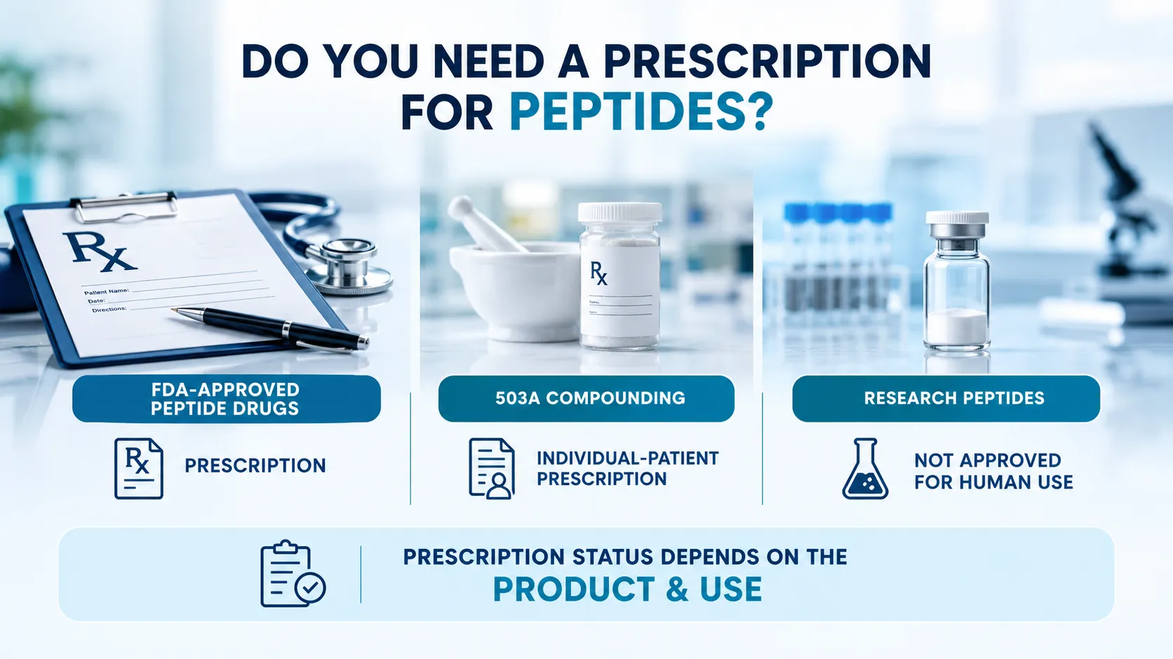
Category 1 in FDA's interim 503A framework refers to nominated substances under evaluation that the agency did not flag for significant safety risk. Thymosin alpha-1 was placed in Category 2, not Category 1, and its removal from Category 2 in 2024 reflected the withdrawal of its nomination rather than a reclassification upward. Removal from Category 2 is not authorization to compound: under Section 503A, a bulk substance must have a USP or NF monograph, be a component of an FDA-approved drug, or appear on the 503A Bulks List. Thymosin alpha-1 meets none of those conditions.
None of this bears on research-use-only materials, which are a separate category entirely — sold as laboratory chemicals, not as drugs. Our explainer on the 503A Bulks List and the 2026 PCAC vote covers the compounding framework in full, and RUO compliance in the USA covers the research-material side.
This article describes published research and regulatory history only. Helix Bio supplies research materials for in-vitro laboratory use. Nothing here is a protocol, a dosing recommendation, or guidance for human or veterinary administration.
What "Dosage Protocol Research" Means Here
Searches for thymosin alpha 1 dosage protocol research usually want one of two things: a schedule to follow, or an understanding of how published studies were designed. Only the second is a legitimate research question, and it is worth answering properly.
Across the international clinical literature, subcutaneous regimens were reported in the range of 1.6 mg administered twice weekly over roughly 26 weeks in the chronic hepatitis B trials, while the TESTS sepsis protocol used 12-hourly administration over seven days. That is a large difference in exposure pattern for the same molecule, and it matters analytically: chronic viral infection studies were designed around sustained immune re-education over months, while critical care studies were designed around acute immune rescue over days. Comparing outcomes across those two designs as if they tested "TA-1" as a single intervention is a category error.
These figures are recorded here as study methodology, in the same way a paper's methods section is cited. They are not recommendations, they do not apply outside the populations and settings studied, and they have no application to laboratory research materials.
Working With TA-1 as a Research Material
For laboratory use, the questions that matter are analytical rather than clinical. TA-1 is supplied as a lyophilized powder and is water-soluble. Because the peptide is acidic and highly charged, identity confirmation by mass spectrometry is particularly useful — the acetylated form differs from the non-acetylated form by 42 Da, a difference that mass spectrometry resolves cleanly and that HPLC purity alone will not tell you.
That makes lot-specific documentation more than a formality for this compound. Reviewing both the HPLC purity figure and the mass-spectrometry identity confirmation on the certificate of analysis is the practical way to confirm you are working with acetylated Tα1 rather than a related species. General handling and reconstitution practice is covered in the peptide reconstitution guide.
For an N-terminally acetylated peptide like Tα1, the mass-spectrometry section of the COA carries information HPLC purity cannot: it confirms the acetyl modification is present, not merely that the material is chemically pure.
Evidence Gaps and Open Questions
Several questions remain genuinely unresolved, and they are more useful to a researcher than another summary of what is already known:
- Does TA-1 bind TLRs directly? Pathway dependence is well demonstrated. Direct receptor engagement, with binding affinities, is far less well characterized.
- Why did TESTS diverge from the pooled small trials? Population differences, standard-of-care differences, and small-study bias are all candidate explanations, and distinguishing them matters for every other immunomodulator in sepsis.
- Is there a definable responder population? The TESTS age and diabetes subgroup signals, and the hepatitis B genotype-response observations, both hint at patient-level heterogeneity that has never been prospectively tested.
- What does the delayed hepatitis B response actually reflect? A durable change in immune control is the proposed explanation, but the mechanism was never directly demonstrated.
- Is the immunosenescence rationale testable? No trial has directly measured naive T-cell output or repertoire diversity as a primary endpoint.
The realistic summary is that thymosin alpha-1 has an unusually deep mechanistic literature, an unusually long international clinical history, a strong tolerability record, and an efficacy picture that becomes less certain as trial quality increases. That combination is uncommon, and it is what makes the TA-1 peptide worth studying carefully rather than describing confidently.
Frequently Asked Questions
Thymosin alpha-1 (Tα1, TA-1) is a 28-amino-acid, N-terminally acetylated peptide corresponding to the first 28 residues of prothymosin alpha, with a molecular weight of approximately 3,108 Da. It was the first peptide isolated from thymic tissue, identified in 1977 from a bovine thymus extract known as thymosin fraction 5. Its synthetic form carries the nonproprietary name thymalfasin.
In published research, TA-1 is studied as an immune-modulating peptide across chronic viral infection models, sepsis and critical care, oncology adjunct settings, and immunosenescence. In laboratory work it is used to investigate T-cell differentiation, dendritic cell maturation, natural killer cell activity, and Toll-like receptor signaling. Research-use-only material is not intended for human or veterinary administration.
Research describes TA-1 acting indirectly rather than by binding T-cells directly: it is reported to prime dendritic cells through TLR-dependent signaling, driving IL-12 production and Th1 polarization, which in turn promotes T-cell differentiation and cytokine output including IL-2 and interferon-gamma. Studies also report upregulation of CD3, CD4 and CD8 markers during thymocyte maturation in research models.
Published findings on CD4+ and CD8+ T-cells span four distinct endpoints — cell counts, activation markers, differentiation state, and functional capacity — and these should not be conflated. Some clinical studies report increased circulating counts, most often in subjects who were lymphopenic at baseline, while other work reports changes in function without changes in number. A general claim that TA-1 simply increases T-cells is stronger than the evidence supports.
They are different peptides from unrelated parent proteins. Thymosin alpha-1 is a 28-amino-acid fragment of prothymosin alpha studied for immune modulation, while TB-500 is a seven-amino-acid fragment of thymosin beta-4 (Ac-LKKTETQ) studied for actin binding, cell migration and tissue repair. The shared word thymosin reflects their common isolation from thymic tissue, not a shared mechanism.
Increased natural killer cell cytotoxic activity is reported across the TA-1 literature, primarily from in vitro and ex vivo cytotoxicity assays. These are laboratory readouts. An increase in measured NK activity in an assay does not by itself establish antiviral or antitumor effect in a living organism, and research framing should preserve that distinction.
The best-supported interactions in the literature involve TLR9 on plasmacytoid dendritic cells, driving MyD88- and IRF7-dependent type I interferon signaling, and TLR2, driving NF-κB and p38 MAPK signaling. Associations with TLR3, TLR4 and TLR7 have also been described, mainly in review literature aggregating results across different systems. Much of this evidence demonstrates pathway dependence in receptor-deficient models rather than direct receptor binding.
No. Thymosin alpha-1 is not approved by the FDA for any indication in the United States. It has held orphan-drug designations, which are development incentives rather than marketing authorizations, and its synthetic form thymalfasin is approved as a prescription medicine in more than 35 other countries. Non-U.S. approval does not confer U.S. approval.
Thymalfasin is the International Nonproprietary Name for the synthetic form of thymosin alpha-1, and Zadaxin is the brand name under which thymalfasin is marketed internationally. All three names refer to the same 28-residue sequence; what differs is regulatory and manufacturing context, not molecular identity.
No. Thymosin alpha-1 was placed in Category 2 of the FDA's interim 503A bulk drug substance policy in September 2023, not Category 1. It was removed from Category 2 on September 20, 2024 after the nomination was withdrawn, reviewed by the Pharmacy Compounding Advisory Committee on December 4, 2024 with FDA proposing against inclusion, and it does not currently appear on the 503A Bulks List. The phrase 'FDA compounding Category 1' is a search term, not an accurate description of its status.
A meta-analysis of five randomized trials covering 353 patients found that thymosin suppressed hepatitis B viral replication with a distinctive delayed pattern: no significant separation at end of treatment, with response rates increasing over 6 to 12 months of follow-up (odds ratio approximately 2.67 at 12 months). A separate meta-analysis of four trials comparing Tα1 with interferon-alpha showed the same shape. These trials were small and predate current nucleos(t)ide analogue standards of care.
TESTS, published in the BMJ in 2025, was a multicentre double-blinded placebo-controlled phase 3 trial in 1,106 adults with sepsis across 22 centres, and it found no significant difference in 28-day all-cause mortality (23.4% versus 24.1% with placebo). Earlier meta-analyses of smaller trials had suggested a mortality benefit. The divergence is a case study in how mechanistic plausibility and pooled small-trial results can fail to survive adequate powering.
- 01Wu J, et al. The efficacy and safety of thymosin α1 for sepsis (TESTS): multicentre, double blinded, randomised, placebo controlled, phase 3 trial. BMJ. 2025.
- 02Efficacy of thymosin α1 for sepsis: a systematic review and meta-analysis of randomized controlled trials. Front Cell Infect Microbiol. 2025.
- 03Li C, Bo L, Liu Q, Jin F. The efficacy of thymosin α1 as immunomodulatory treatment for sepsis: a systematic review of randomized controlled trials. BMC Infect Dis. 2016;16:488.
- 04Chan HL, et al. The efficacy of thymosin in the treatment of chronic hepatitis B virus infection: a meta-analysis. Aliment Pharmacol Ther. 2001.
- 05Comparison of the efficacy of thymosin alpha-1 and interferon alpha in the treatment of chronic hepatitis B: a meta-analysis. Antiviral Res. 2008.
- 06Tao N, et al. Thymosin α1 and its role in viral infectious diseases: the mechanism and clinical application. Molecules. 2023;28(8):3539.
- 07Romani L, et al. Thymosin α1 activates the TLR9/MyD88/IRF7-dependent murine cytomegalovirus sensing for induction of anti-viral responses in vivo. Int Immunol. 2007;19(11):1261-1270.
- 08U.S. Food and Drug Administration. Pharmacy Compounding Advisory Committee Meeting, December 4, 2024 — Voting Questions (thymosin alpha-1 free base and acetate).
- 09U.S. Food and Drug Administration. Thymosin Alpha-1 (Tα1) Related Bulk Drug Substances — PCAC presentation, December 4, 2024.
Helix Bio Chem Team
Research & Product Team
Our in-house team tracks published peptide research and translates it into clear, source-cited summaries for the research community.
Reviewed by in-house research chemists
support@helixbiochem.com