LL-37 Peptide Research: Human Cathelicidin, hCAP18 & Mechanisms
Recovery protocolsSeptember 10, 202617 min read
LL-37 is the only human cathelicidin, released from hCAP18 by proteolysis. What its sequence, membrane mechanism and two human trials actually establish.
LL-37 is the only cathelicidin antimicrobial peptide identified in humans, while cathelicidin itself is a family name spanning many mammalian species.
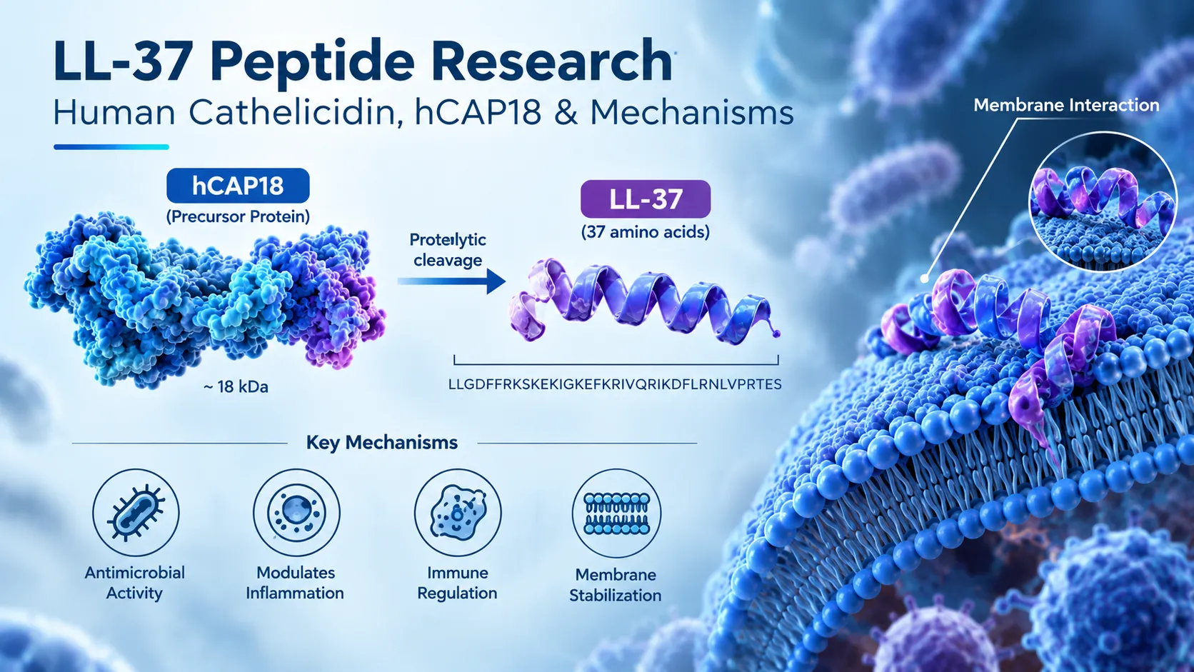
LL-37 is the 37-residue C-terminal peptide released from the precursor hCAP18, which is encoded by the CAMP gene on chromosome 3p21.31.
In neutrophils, proteinase 3 cleaves hCAP18 to release LL-37, but skin and seminal plasma proteases generate different peptides from the same precursor.
The verified LL-37 sequence is LLGDFFRKSKEKIGKEFKRIVQRIKDFLRNLVPRTES, corresponding to UniProt P49913 residues 134 to 170, with an average molecular weight near 4,493 Da.
LL-37 carries a net charge of approximately +6 and folds into an amphipathic alpha-helix on anionic surfaces, which is the basis of its selectivity for bacterial membranes.
FPR2-mediated chemotaxis is a demonstrated receptor interaction; EGFR effects appear to be indirect transactivation and P2X7 involvement is described as modulation rather than direct binding.
By complexing extracellular self-DNA and self-RNA and delivering them to endosomal TLR9 and TLR7/8, LL-37 contributes to psoriasis pathophysiology, so its biology is context-dependent rather than uniformly protective.
The 148-patient phase IIb HEAL LL-37 trial did not meet its primary endpoint of complete wound closure; a positive finding came only from a post hoc subgroup the trial was not powered to test.
Cathelicidin LL-37 was removed from FDA 503A Category 2 effective 22 April 2026 because the nomination was withdrawn, not because of a safety finding, and a PCAC consultation is scheduled before the end of February 2027.
LL-37 has no tryptophan or tyrosine, so absorbance at 280 nm cannot determine peptide content and chromatographic analysis must run in the low-UV region.
LL-37 occupies an unusual position in peptide research. It is not a designed analogue or a fragment someone excised from a larger hormone to see what would happen. It is a molecule the human body already makes, in neutrophils and epithelial cells, and it has been characterised in thousands of peer-reviewed papers since 1995. That depth of literature is exactly why the compound is easy to write about badly. There is enough published work to support almost any claim if you select carefully, and enough mechanistic detail to make a page sound authoritative without ever specifying which experimental system produced the result.
Featured In This Article
LL-37
RESEARCH PEPTIDE
Highly purified synthetic peptide prepared for rigorous laboratory research.
$77.00
This article takes the opposite approach. It covers what LL-37 is, where it comes from at the gene level, what its sequence and structure actually explain, how it interacts with bacterial membranes, what the receptor evidence supports and what it only suggests, and — the part most pages skip — what happened when synthetic LL-37 was put into humans. Two controlled trials exist. One is usually cited as a success and the other as a success too, which is not what the published data say.
What Is LL-37?
LL-37 is the mature antimicrobial peptide released from the C-terminal end of hCAP18, the precursor protein encoded by the human CAMP gene. It is 37 amino acids long, carries a net positive charge of roughly +6 at physiological pH, contains no cysteine, and folds into an amphipathic alpha-helix on contact with anionic surfaces. It is the only cathelicidin identified in humans.
That last point resolves the most common confusion in this topic. Cathelicidin is a family name, not a molecule name. Many mammals carry several cathelicidin genes — pigs have a whole set, including PR-39 and the protegrins; cattle have the bactenecins. Humans have exactly one cathelicidin gene, and its product is LL-37. So "human cathelicidin" and "LL-37" refer to the same peptide, while "cathelicidin" on its own refers to a class spanning many species. When a paper says "cathelicidin" and means the mouse molecule, it is talking about CRAMP, which is the murine orthologue and is not sequence-identical to LL-37 — a distinction that matters whenever a rodent result gets carried into a human claim.
LL-37 and the Human Cathelicidin System
From CAMP Gene to hCAP18
The CAMP gene sits on chromosome 3p21.31 and contains four exons. It encodes a 170-residue prepropeptide with three parts: an N-terminal signal sequence, a conserved cathelin-like domain that gives the family its name, and the C-terminal antimicrobial domain. Removing the signal peptide yields the 18 kDa proform, hCAP18 — the "18" is its apparent molecular weight, not a residue count.
hCAP18 is not an inert storage form. It is packaged into the specific (secondary) granules of neutrophils and is also expressed by keratinocytes, mucosal and airway epithelium, monocytes, macrophages, mast cells and lymphocytes. Transcription of CAMP is inducible, and one of the better-characterised inducers is the active form of vitamin D acting through the vitamin D receptor — which is why circulating hCAP18 and 25-hydroxyvitamin D levels have been examined together in human observational studies.
How hCAP18 Becomes LL-37
The precursor has to be cut. In neutrophils, the enzyme responsible is proteinase 3, a serine protease that cleaves hCAP18 extracellularly after granule exocytosis to liberate the 37-residue peptide.
It would be tidy if that were the whole story, but it is not, and the untidiness is scientifically useful. Processing is site-dependent. On the skin surface, the CAMP gene product is handled by a serine-protease-dependent mechanism that generates several shorter peptides distinct from LL-37 — KR-20, RK-31, KS-30, LL-23, LL-29 and FF-33 among them, each corresponding to a different residue span of the precursor. These fragments are not degradation debris; they have their own antimicrobial profiles, and some retain activity under salt conditions where LL-37 itself is inhibited. In seminal plasma, the precursor is cleaved by gastricsin under acidic conditions to yield a 38-residue peptide, ALL-38, which differs from LL-37 by a single N-terminal alanine.
The practical consequence is that "cathelicidin activity" measured in a tissue is not necessarily LL-37 activity. It may be a mixture whose composition depends on which proteases are present.
Entity
What it is
Relationship
CAMP gene
Single human cathelicidin gene, chromosome 3p21.31, four exons
Encodes the 170-residue prepropeptide
Prepropeptide
Signal sequence + cathelin-like domain + C-terminal antimicrobial domain
Signal removal yields hCAP18
hCAP18
18 kDa proform stored in neutrophil specific granules
Product of hCAP18 cleavage by proteinase 3 in neutrophils
KR-20, RK-31, ALL-38 and others
Alternative cleavage products of the same precursor
Generated by tissue-specific proteases, not by proteinase 3
Why It Is Called LL-37
The name is a fossil of a correction, and the correction is worth knowing.
When the human sequence was first deduced from bone marrow cDNA in 1995, the investigators predicted where the antimicrobial domain would begin and named the putative peptide FALL-39 — after the first four predicted residues, Phe-Ala-Leu-Leu, and a predicted length of 39. The naming followed the convention already used for the porcine cathelicidin PR-39. When the peptide was subsequently isolated from human granulocytes rather than predicted from sequence, the real mature form turned out to start two residues later, at a pair of leucines, and to be 37 residues long. It was renamed LL-37 accordingly.
So the name encodes an empirical finding: two leading leucines, 37 residues. FALL-39 still appears in older literature and in some antibody catalogues, where it denotes residues 132-170 rather than 134-170.
Sequence, Structure and Molecular Identity
The verified sequence is LLGDFFRKSKEKIGKEFKRIVQRIKDFLRNLVPRTES, corresponding to residues 134-170 of UniProt entry P49913.
Read the composition rather than just the letters. There are eleven basic residues — six lysines and five arginines — against five acidic ones, giving the net positive charge that drives everything downstream. There is no cysteine at all, so LL-37 has no disulfide constraint and no oxidation liability at sulphur. There is no tryptophan and no tyrosine either, which has an analytical consequence covered later on this page.
In dilute salt-free water the peptide is largely disordered. It becomes helical in the presence of anions, on membrane surfaces, and at higher ionic strength, and the degree of helicity tracks with activity. The helix is amphipathic: hydrophobic residues cluster on one face, cationic residues on the other. Structural work describes a rigid helical region stabilised in part by a salt bridge between Glu16 and Lys12 and a hydrophobic cluster involving Ile13, Phe17 and Ile20, with a flexible C-terminal tail.
Property
Value
Source / verification
Length
37 residues
UniProt P49913, residues 134-170
Sequence
LLGDFFRKSKEKIGKEFKRIVQRIKDFLRNLVPRTES
UniProt P49913; consistent across peer-reviewed reviews
Average molecular weight
Approximately 4,493 Da
Calculated from the 134-170 sequence
Net charge at neutral pH
Approximately +6
Six Lys and five Arg against five acidic residues
Cysteine content
None
Reported as cysteine-free in the original 1995 characterisation
Aromatic residues
Four phenylalanine; no tryptophan or tyrosine
A note on identifiers: CAS numbers and database compound IDs circulate for LL-37, but they are applied inconsistently across sources — some to the free peptide, some to salt forms, some to the precursor. They are omitted here rather than reproduced without a single authoritative anchor. UniProt P49913 residues 134-170 is the identity reference that resolves unambiguously.
How LL-37 Interacts With Bacterial Membranes
The short answer is electrostatics first, hydrophobicity second, and the outcome depends on the membrane.
Bacterial cytoplasmic membranes present a substantially anionic outer surface — phosphatidylglycerol and cardiolipin in Gram-positive organisms, and in Gram-negative bacteria an outer membrane whose lipopolysaccharide layer carries phosphate and carboxylate groups. Mammalian plasma membranes present mostly zwitterionic phosphatidylcholine outward, with the anionic phospholipids sequestered on the inner leaflet. A peptide carrying +6 therefore encounters a charge gradient that favours microbial surfaces before any structural change occurs.
Once bound, the induced amphipathic helix buries its hydrophobic face in the acyl chain region. What happens next is where careful writing matters. Model membrane studies describe LL-37 accumulating on the surface and, above a threshold surface concentration, permeabilising the bilayer through a carpet-like mechanism rather than forming the well-defined, long-lived barrel-stave channels that some other peptides make. Transient toroidal pores have been described, and the two descriptions are not mutually exclusive — they can represent different regimes of the same concentration-dependent process.
LL-37 also binds lipopolysaccharide directly and neutralises its capacity to trigger inflammatory signalling. That is a separate activity from killing: LPS neutralisation blunts the host response to bacterial material whether or not the bacterium is alive.
Why Conditions Change the Answer
Three variables recur throughout the microbiology literature and should qualify any statement about LL-37 potency:
Ionic strength. Cations compete for the anionic sites the peptide relies on. Activity measured in low-salt buffer is not activity in physiological saline.
Serum and protein. Serum components bind LL-37 and reduce free peptide concentration, which is one reason in vitro potency does not extrapolate to systemic settings.
Organism and growth state. Susceptibility differs between Gram-positive and Gram-negative species, and biofilm-resident cells behave differently from planktonic ones.
There is also a boundary that vendor pages rarely state: the concentration range in which LL-37 kills bacteria in vitro is not comfortably below the range in which it damages mammalian cells. The two windows overlap. Reviews of LL-37 as a therapeutic candidate consistently name cytotoxicity, alongside proteolytic instability and manufacturing cost, as a principal obstacle — which is precisely why so much of the field works on engineered analogues rather than the native sequence.
The Second Arm: Immunomodulation and Receptor Signalling
Direct killing is only half the biology, and arguably not the more consequential half at physiological concentrations. LL-37 also acts on host cells, and here the evidence needs grading rather than listing.
FPR2 (formyl peptide receptor 2, formerly FPRL1) is the best-supported receptor interaction. LL-37 was shown to chemoattract human neutrophils, monocytes and T cells through this G-protein-coupled receptor in work published in the Journal of Experimental Medicine in 2000, and FPR2-dependent responses have since been described in angiogenesis and epithelial contexts. This is a demonstrated receptor-mediated effect, not an inference.
EGFR transactivation is the mechanism most often invoked for LL-37's effects on epithelial cells and keratinocytes — cytokine release, migration, re-epithelialisation. The evidence supports indirect activation, typically through metalloproteinase-mediated shedding of an EGFR ligand, rather than LL-37 binding EGFR itself. Some groups have failed to reproduce downstream ERK activation in their systems, so this belongs in the "supported but not uniform" column.
P2X7 appears in the literature as a modulator of LL-37 responses in macrophages, particularly around ATP-driven inflammasome signalling. The reports describe modulation of the receptor's response rather than a demonstration of direct high-affinity binding, and much of it comes from murine cell lines.
Toll-like receptors are where LL-37's role is clearest and least comfortable. LL-37 does not act as a conventional TLR agonist. It acts as a carrier: it binds extracellular nucleic acids released by dying cells, protects them from degradation, condenses them into aggregates and delivers them into endosomal compartments where TLR9 and TLR7/8 reside. Complexed with self-DNA it drives interferon-alpha production by plasmacytoid dendritic cells; complexed with self-RNA it additionally activates myeloid dendritic cells.
Evidence grading used on this page: **demonstrated** means a direct experimental result in a defined system; **supported** means multiple independent reports with a consistent mechanism; **proposed** means a model consistent with the data but not established. FPR2 chemotaxis is demonstrated. EGFR transactivation is supported. Direct P2X7 binding is proposed.
When LL-37 Is Not Protective
The nucleic-acid-carrier mechanism above is not a curiosity. It is the accepted molecular explanation for how LL-37 contributes to psoriasis, established in a 2007 Nature paper and extended since. LL-37 is also a T-cell autoantigen in psoriasis, with LL-37-reactive CD4 and CD8 cells detectable in patient circulation and correlating with disease activity. LL-37-reactive T cells and anti-LL-37 antibodies have separately been described in systemic lupus erythematosus. Abnormal cathelicidin processing is central to current models of rosacea pathophysiology.
None of that contradicts the antimicrobial biology. It means the same molecule has different consequences depending on concentration, tissue, what else is in the extracellular space, and which cells are present to respond. A peptide that condenses free DNA is useful when the DNA came from a pathogen and harmful when it came from the host's own dying keratinocytes. Any page that presents LL-37 as uniformly beneficial has simply not read the dermatology literature.
What the Evidence Shows, Model by Model
Evidence type
Model
Question studied
Main finding
Principal limitation
Biophysical
Synthetic lipid vesicles, monolayers
How does LL-37 perturb a bilayer?
Surface accumulation then concentration-dependent permeabilisation; amphipathic helix induced on binding
Model membranes lack outer membrane, wall, proteins and active repair
In vitro microbiology
Bacterial and fungal cultures
Which organisms are susceptible?
Broad-spectrum activity across Gram-positive and Gram-negative species; LPS binding and neutralisation
Strongly modulated by salt, serum and growth state; killing and cytotoxicity windows overlap
Cell culture
Neutrophils, monocytes, keratinocytes, epithelial and dendritic cells
The Human Trials, Read Properly
Two controlled trials of synthetic LL-37 in humans have been published. Both were topical applications to venous leg ulcers, both were run by the same Swedish sponsor, and both used the acetate salt of the peptide in a polyvinyl alcohol vehicle. There is no published trial of injected or systemically administered LL-37.
The first-in-man study (Grönberg and colleagues, Wound Repair and Regeneration, 2014) enrolled 34 participants. After a three-week placebo run-in, patients received twice-weekly applications of LL-37 at one of three concentrations, or placebo, for four weeks. The healing rate constant was about sixfold higher than placebo at the lowest concentration, with a p-value of 0.003. At the intermediate concentration the effect was about threefold but did not reach significance. At the highest concentration there was no improvement at all, and local reactions at the wound site were more frequent and more severe.
That is not a dose–response curve. It is an inverted one, and only one of three arms was statistically significant.
The phase IIb study (Mahlapuu and colleagues, Wound Repair and Regeneration, 2021) tested this properly: 149 patients randomised across 15 sites in Poland and Sweden, 148 treated, thirteen weeks of twice-weekly treatment, with confirmed complete wound closure as the primary endpoint. The highest concentration was dropped on the basis of the first trial.
The primary endpoint was not met. Estimated complete closure was 26.5% at the low concentration, 24.7% at the higher one, and 25.3% on placebo. No secondary endpoint reached significance in the full population either.
A post hoc analysis restricted to patients whose wounds were at least 10 cm² at randomisation did find significant differences favouring the low concentration — 28.1% closure against 8.1% for placebo, odds ratio 4.454, p = 0.0458, with consistent movement in several related healing measures. The authors report this as an interesting observation and state plainly that the trial was not designed or powered to test subgroups, that the subgroups contained only 21 to 24 patients per arm, and that an adequately powered study is needed. They also note that wound-area measurement is less precise for small ulcers, which could contribute to the split.
A post hoc subgroup finding in a trial that missed its primary endpoint is hypothesis-generating, not evidence of efficacy. LL-37 has not demonstrated clinical benefit in a prespecified analysis of a controlled trial in any indication, and there is no approved LL-37 product anywhere.
Tolerability was the more solid finding. Across both trials, at the concentrations carried forward, local application was well tolerated, with no deaths and no serious adverse events attributed to the study drug in the phase IIb.
Where LL-37 Sits With the FDA
This is the section most likely to be misread, so it is worth being exact about what changed in 2026.
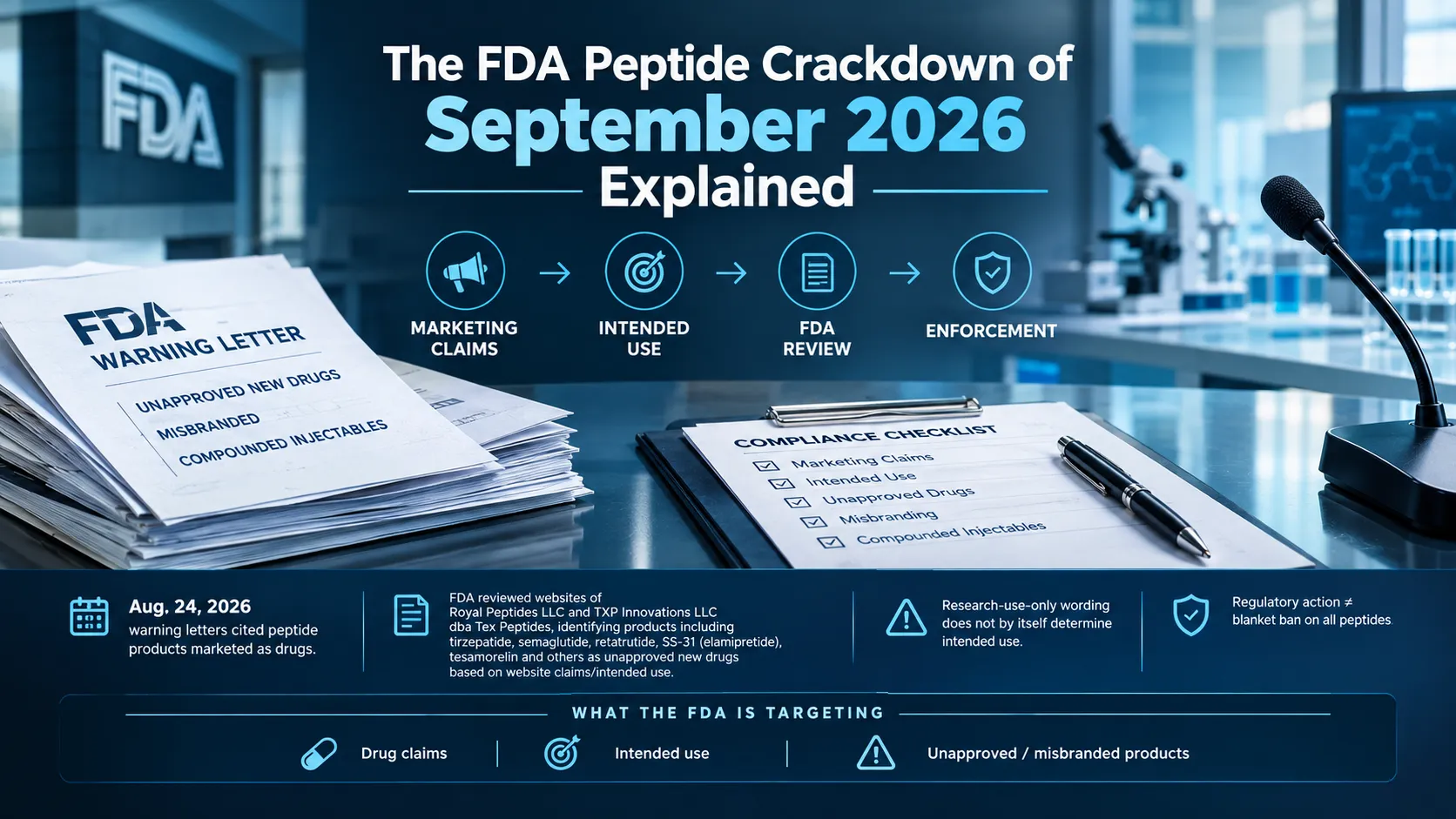
Cathelicidin LL-37 was placed in Category 2 of the FDA's interim 503A bulk drug substances list in September 2023 — the grouping for substances the agency considered to raise significant safety concerns, citing immunogenicity risk, impurity and characterisation questions, and limited human clinical data. Effective 22 April 2026, LL-37 was removed from Category 2, together with eleven other peptides. The stated reason was that the nominations had been withdrawn by the nominators. No new safety or efficacy data accompanied the change.
LL-37 was not among the seven peptides reviewed by the Pharmacy Compounding Advisory Committee on 23–24 July 2026. It is one of five substances — with injectable GHK-Cu, Melanotan II, dihexa acetate and PEG-MGF — slated for a separate committee consultation before the end of February 2027.
Issue
Current status
What it means
What it does NOT mean
Category 2 removal
Removed effective 22 April 2026
The nomination was withdrawn, so FDA stopped evaluating it under that nomination
It does not mean FDA found the substance safe
Current category
In none of the three categories
It sits outside the Category 1 enforcement-discretion policy as well
It is not on the 503A bulks list and not in Category 1
PCAC review
Consultation scheduled before end of February 2027
An advisory committee will consider whether to recommend inclusion
Scheduling a review is not a decision, and the committee only advises
FDA approval
None
Two further points. First, in each of the twelve cases FDA's own scientists had recommended against inclusion, citing insufficient safety, efficacy and moiety characterisation information — the committee's July votes went the other way on six of seven, and the committee only advises. Second, even a favourable recommendation would still require notice-and-comment rulemaking before anything is added to the list. If you follow this area, our explainer on the 2026 503A bulks list vote sets out the mechanics.
Analytical Notes for LL-37 Research Material
Three characteristics of this particular sequence change how you verify a batch.
It has no useful 280 nm chromophore. LL-37 contains four phenylalanines and neither tryptophan nor tyrosine, so its extinction coefficient at 280 nm is effectively zero. A280 cannot be used to determine peptide content. This is not theoretical: the drug product used in the phase IIb trial had its content and impurities determined by liquid chromatography with UV detection at 217 nm, in the backbone absorbance region, with identity confirmed by LC-MS. Any purity figure for LL-37 should have been generated by a low-UV method.
Purity and content are different numbers. An area-percent chromatographic figure describes the proportion of detected material that is the target peak. It says nothing about how much of the vial's mass is peptide as opposed to counterion, residual water and salt. LL-37 is typically supplied as an acetate salt, and a highly charged 37-mer carries a substantial counterion load. Our guide to reading a certificate of analysis covers where these two figures appear and how they differ.
It is cationic, amphipathic and adsorptive. Peptides with this profile bind to glass and plastic surfaces and can aggregate at air–liquid interfaces, which means apparent concentration in a working solution may not match nominal concentration. Vehicle composition, ionic strength and pH also determine how much of the peptide is helical, and helicity tracks with activity — so the same material can behave differently in two buffers.
Because LL-37's conformation is buffer-dependent, an activity result is only interpretable alongside the exact solution conditions it was measured in. Record ionic strength, pH and protein content with every assay, not just peptide concentration.
Helix Bio supplies LL-37 as a lyophilised research peptide with HPLC and mass spectrometry verification and a certificate of analysis per batch, for laboratory research use only. High chromatographic purity confirms what is in the vial; it does not establish biological activity, and a certificate of analysis is a manufacturing document, not evidence of clinical effect.
What the Evidence Establishes, and What It Does Not
Established: LL-37 is the sole human cathelicidin; it is released from hCAP18 by proteinase 3 in neutrophils and by other proteases elsewhere; its sequence, charge and inducible helical structure are well characterised; it permeabilises anionic membranes in vitro and binds LPS; it chemoattracts leukocytes through FPR2; and it delivers extracellular nucleic acids to endosomal TLRs, which is how it participates in psoriasis.
Not established: that in vitro antimicrobial potency predicts behaviour in an infected human tissue; that any receptor interaction beyond FPR2 involves direct binding; that LL-37 improves wound healing in a controlled human trial with a prespecified endpoint; and that its biology is beneficial independent of context.
That gap between mechanism and outcome is the honest summary of this compound. It is one of the best-characterised antimicrobial peptides in biology and one of the least translated. For adjacent immune-signalling compounds where the evidence has a different shape, KPV offers a useful contrast — a three-residue fragment with a defined transport mechanism and a much thinner literature — as do thymosin alpha-1, which has an actual approved-product history outside the US, and ARA-290, which reached four phase 2 trials on a receptor model that is still contested.
Got Questions?
Frequently Asked Questions
Not quite — LL-37 is the only cathelicidin humans have, but cathelicidin is a family name covering many species. Pigs, cattle, mice and other mammals carry cathelicidin genes producing different peptides, so "cathelicidin" alone does not identify a molecule. When a paper refers to human cathelicidin it means LL-37; when it refers to mouse cathelicidin it means CRAMP, which is the orthologue but not sequence-identical.
hCAP18 is the 18 kDa precursor protein and LL-37 is the mature peptide cut from its C-terminal end. hCAP18 consists of a conserved cathelin-like domain plus the antimicrobial domain; proteolytic cleavage separates them and releases the 37-residue active peptide. Older papers write "hCAP18/LL-37" when they are measuring the precursor and the mature peptide together, which is common in tissue and plasma studies.
The sequence is LLGDFFRKSKEKIGKEFKRIVQRIKDFLRNLVPRTES, corresponding to residues 134 to 170 of UniProt entry P49913. It contains six lysines and five arginines against five acidic residues, giving a net charge near +6, and it contains no cysteine, no tryptophan and no tyrosine. Its average molecular weight is approximately 4,493 Da.
Because the mature peptide begins with two leucines and is 37 residues long — and the name records a correction. When the human sequence was first deduced from cDNA in 1995 the antimicrobial domain was predicted to start four residues earlier and was named FALL-39 after the predicted residues Phe-Ala-Leu-Leu. Isolation from granulocytes showed the real mature form started two residues further along, so it was renamed LL-37.
In neutrophils, proteinase 3 cleaves hCAP18 extracellularly after granule release to produce LL-37. That is not the only route, though. On skin, serine-protease-dependent processing generates shorter peptides such as KR-20, RK-31 and LL-23 instead, and in seminal plasma gastricsin produces a 38-residue form called ALL-38 under acidic conditions. Which peptide you get depends on the tissue and its proteases.
Through charge-driven binding followed by membrane permeabilisation. The peptide's net positive charge attracts it to the anionic surfaces of bacterial membranes and lipopolysaccharide, it folds into an amphipathic helix on contact, and above a threshold surface concentration it disrupts the bilayer — described in model systems as a carpet-like mechanism with transient pore formation rather than stable channels. Potency depends heavily on salt concentration, serum proteins and the organism tested.
No. Activity has been reported across Gram-positive and Gram-negative bacteria, fungi and enveloped viruses, but susceptibility varies by organism, growth state and assay conditions, and the mechanisms differ. Biofilm-resident bacteria behave differently from planktonic cultures, and lipopolysaccharide neutralisation is a distinct activity from killing — it reduces the host inflammatory response to bacterial material whether or not the organism dies.
FPR2, the formyl peptide receptor 2, is the best-supported direct interaction and mediates chemotaxis of neutrophils, monocytes and T cells. Effects attributed to EGFR appear to run through indirect transactivation rather than LL-37 binding EGFR itself, and reports involving P2X7 describe modulation of the receptor's response rather than demonstrated direct binding. LL-37's relationship with Toll-like receptors is different again: it carries nucleic acids into endosomes rather than acting as a receptor agonist.
No, and this is one of the clearer findings in the field. By binding extracellular self-DNA and self-RNA and delivering them to endosomal TLR9 and TLR7/8, LL-37 drives interferon production by dendritic cells — the accepted mechanism for its contribution to psoriasis, established in a 2007 Nature study. LL-37 is also a recognised T-cell autoantigen in psoriasis, and altered cathelicidin processing features in rosacea models. The same molecular property is protective against pathogens and problematic in chronic inflammation.
Two published randomised controlled trials, both topical applications to venous leg ulcers. The 34-patient first-in-man study reported significantly faster healing at only the lowest of three concentrations, with no benefit and more local reactions at the highest. The 148-patient phase IIb study did not meet its primary endpoint of confirmed complete wound closure; a post hoc subgroup of patients with wounds of at least 10 cm² showed significant differences, which the authors describe as hypothesis-generating. No trial of injected or systemic LL-37 has been published.
LL-37 is not FDA-approved for any indication. It was removed from Category 2 of the interim 503A bulk drug substances list effective 22 April 2026 because the nomination was withdrawn, not because FDA made a safety finding — and removal did not place it in Category 1 or on the bulks list, so it currently sits in no category at all. A Pharmacy Compounding Advisory Committee consultation is scheduled before the end of February 2027. That committee advises; it does not decide, and rulemaking would still follow.
Because the sequence contains no tryptophan and no tyrosine, so its absorbance at 280 nm is effectively zero and A280 cannot determine peptide content. Chromatographic analysis has to run in the low-UV backbone region instead — the LL-37 drug product used in the published phase IIb trial was quantified by liquid chromatography with UV detection at 217 nm, with identity confirmed by LC-MS. A purity figure for LL-37 that cites 280 nm detection should prompt a question about the method.